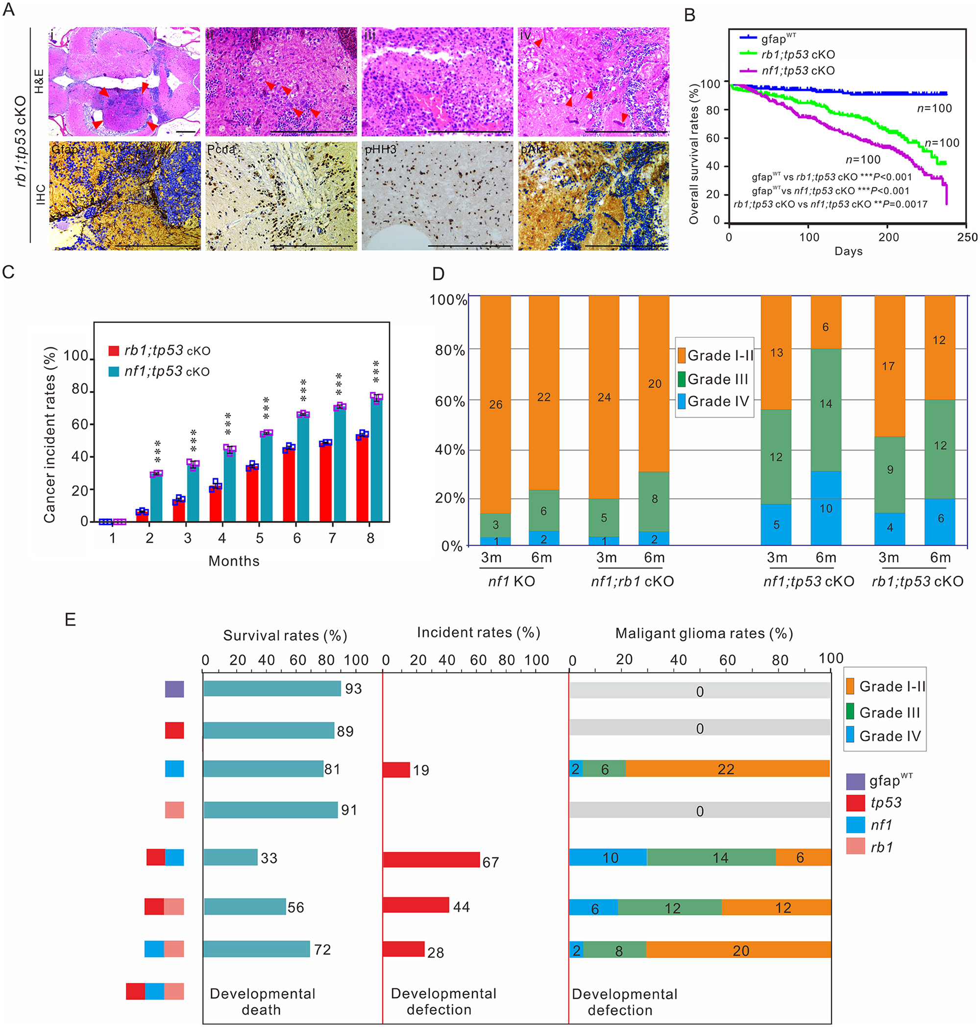
Fig. 5 Tp53 mutation is critical for poor prognosis in zebrafish. (A) Histological examination of gliomas in 3-month-old rb1;tp53 cKO fish. (i–iv) Haematoxylin and eosin (H&E) staining showed the generated tumours (arrowheads; i), multinucleated giant cells (arrowheads; ii), the typical gliomatosis phenotype (iii), and vascularity (arrowheads; iv). Immunohistochemistry staining was performed to examine tumour-relevant indicators, including Gfap, Pcna, pHH3, and pAkt, in brain tissues of rb1;tp53 cKO fish. Scale bars = 100 μm. (B and C) Overall survival rates and cancer incidences of rb1;tp53 cKO and nf1;tp53 cKO fish, respectively (n = 100 for each group). The glioma formation was preliminarily estimated based on the ‘bending-body’ phenotype, and confirmed by haematoxylin and eosin staining. (D) Malignancy of the tumours derived from the fish with various mutations at 3 or 6 months of age (n = 30 for each group). (E) Summary of the survival rates, cancer incidences, and malignancy of the generated gliomas in fish lines with various mutations at 6 months of age. Data shown as mean ± SEM. ***P < 0.001.
Image
Figure Caption
Figure Data
Acknowledgments
This image is the copyrighted work of the attributed author or publisher, and
ZFIN has permission only to display this image to its users.
Additional permissions should be obtained from the applicable author or publisher of the image.
Full text @ Brain

