
|
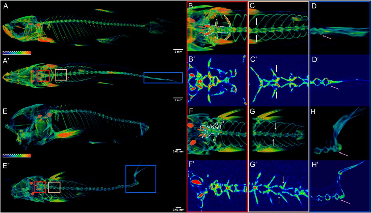
Fig. 2 nkx3.2 mutant zebrafish develop axial skeletal abnormalities. (A,E) µCT imaging of 60 dpf wild types (A,A′) and nkx3.2ua5011/ua5011 mutants (E,E′) shown with gradient 3D rendering in sagittal (A,E) and dorsal (A′,E′) views. (B-D,F-H) Magnifications of three spinal regions: anterior vertebrae (red box, inclusive of the occiput and Weberian apparatus: B,B′,F,F′), precaudal vertebrae with rib articulations (tan box, white arrows indicate corresponding left and right ribs: C,C′,G,G′), and caudal fin vertebrae (blue box, pink arrow indicates caudal-most vertebra: D,D′,H,H′) shown in 3D (B,C,D,F,G,H) and 2D (B′,C′,D′,F′,G′,H′) slice. Scale bars: 1 mm (A,A′); 0.61 mm (E,E′).