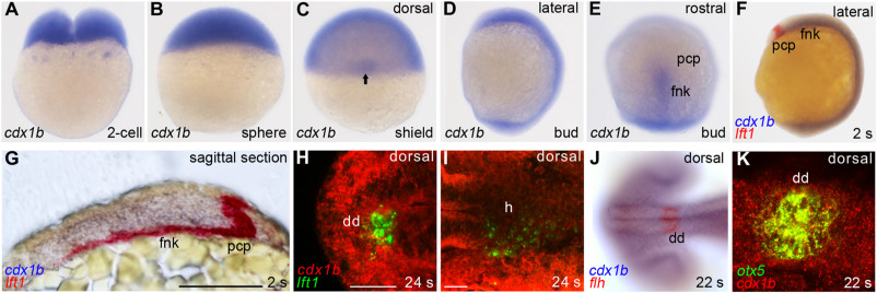
Fig. 1 Fig. 1. cdx1b expression during gastrulation and segmentation stages. RNA in situ hybridization was conducted on embryos from 2-cell (A), sphere (B), shield (C), and bud (D, E) stages using a cdx1b antisense RNA probe. Enriched cdx1b expression in the shield (C, arrow) and forebrain neural keel (E) is shown. Double in situ hybridization was performed on embryos from 2 s (F) stage using cdx1b (blue) and lft1 (red) RNA probes as well as 24 s (H, I) stage using cdx1b (red) and lft1 (green) RNA probes. A sagittal section of stained embryos (G) is shown. Double in situ hybridization was conducted on 22 s embryos with cdx1b (blue) and flh (red) probes (J) or with cdx1b (red) and otx5 (green) RNA probes (K). Scale bar (G) represents 100 μm; scale bar (H, I) represent 40 μm dd, diencephalon; fnk, forebrain neural keel; h, heart; pcp, prechordal plate.
Reprinted from Developmental Biology, 470, Wu, C.S., Lu, Y.F., Liu, Y.H., Huang, C.J., Hwang, S.L., Zebrafish Cdx1b modulates epithalamic asymmetry by regulating ndr2 and lft1 expression, 21-36, Copyright (2020) with permission from Elsevier. Full text @ Dev. Biol.

