IMAGE

Fig 1

ID
ZDB-IMAGE-210113-118
Source
Figures for Nunley et al., 2020
Image
Figure Caption

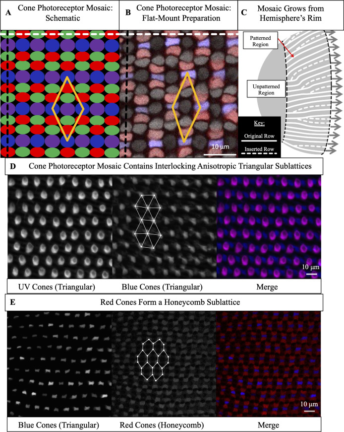
Fig 1 The cone mosaic is composed of four interpenetrating sublattices: two triangular sublattices and two honeycomb sublattices.

(A) Schematic of cone photoreceptors (colored by subtype) in apical plane of zebrafish retina. The ‘unit cell’ (yellow parallelogram) contains one UV cone, one Blue cone, two Green cones, and two Red cones. White dashed line: ‘row’ axis. Black dashed line: ‘column’ axis. (B) Cone mosaic from flat-mount retinal preparation of an adult, triple transgenic fish, Tg[sws2:GFP; trβ2:tdTomato; gnat2:CFP]. Blue cones express a fluorescent reporter (pseudo-colored blue) under control of the sws2 promoter, and Red cones express a fluorescent reporter (pseudo-colored red) under control of the trβ2 promoter. All cones express an additional reporter (pseudo-colored gray) under control of the gnat2 promoter. We distinguish between UV and Green cone subtypes based on morphology. (C) Schematic of photoreceptor epithelium, lining the outer surface of the hemispheric retina. The central retina, which surrounds the hemispheric pole and forms during the larval period, is unpatterned. As the retina grows by mitotic addition of new photoreceptors (and other retinal cells) at the hemispheric rim (gray arrows), there is a disorder-to-order transition (black dashed line). After this transition, the cone mosaic grows by neurogenesis at the hemispheric rim throughout the fish’s life. Because the hemispheric circumference grows, rows of cells are inserted to maintain approximately constant cell spacing. (D) UV and Blue cones in flat-mount retinal preparation from a double transgenic (Tg[sws1:GFP; sws2:mCherry]) line in which UV and Blue cones express distinct fluorescent reporters. UV cones (pseudo-colored magenta) form an anisotropic triangular sublattice that interpenetrates with an anisotropic triangular sublattice of Blue cones (pseudo-colored blue). We connect (white lines) a subset of nearest neighbors in the Blue cone sublattice. (E) Blue (pseudo-colored blue) and Red (pseudo-colored red) cones in flat-mount retinal preparation from panel B. Red cones neighbor Blue cones in each column. The Red cones form an anisotropic honeycomb sublattice. We connect (white lines) a subset of nearest neighbors in the Red cone sublattice; note the different nearest neighbor patterns in the Blue cone triangular sublattice (panel D) and the Red cone honeycomb sublattice (panel E). The Green cones form a honeycomb sublattice (not shown here).

Acknowledgments
This image is the copyrighted work of the attributed author or publisher, and ZFIN has permission only to display this image to its users. Additional permissions should be obtained from the applicable author or publisher of the image. Full text @ PLoS Comput. Biol.