IMAGE

FIGURE 1

ID
ZDB-IMAGE-201211-7
Source
Figures for van Wijk et al., 2020
Image
Figure Caption

FIGURE 1

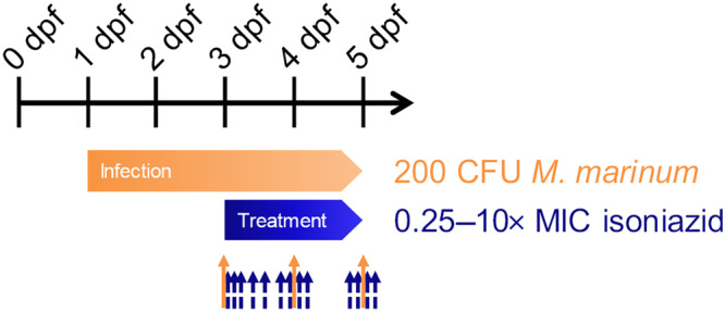
Experimental study design. Fertilized eggs at 0 days post fertilization (dpf) were harvested and injected with 200 CFU Mycobacterium marinum at 1 dpf. After 2 days of establishing the infection, fluorescence imaging was performed (orange arrow; n ≥ 20 larvae per group), and the treatment with isoniazid dissolved in the external treatment medium (0.25–10‐fold MIC, MIC = 15 mg·L−1, and control) was started. Fluorescence imaging was repeated daily (orange solid arrows). In satellite larvae groups, destructive homogenate and blood samples for internal isoniazid exposure quantification were taken from 0 to 50 h of treatment (blue dashed arrows)

Acknowledgments
This image is the copyrighted work of the attributed author or publisher, and ZFIN has permission only to display this image to its users. Additional permissions should be obtained from the applicable author or publisher of the image. Full text @ Br. J. Pharmacol.