Fig. 5
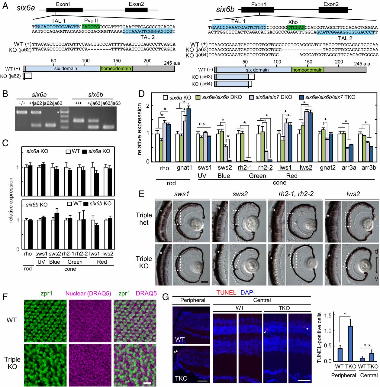
Fig. 5 Compromised development of cone photoreceptors in six6a/six6b/six7 TKO zebrafish. (A) Exon/intron organization and partial nucleotide sequences of zebrafish six6a and six6b. The binding sites of the left and right TAL effector nucleases are highlighted in blue. The recognition sites of the restriction endonucleases PvuII and XhoI are highlighted in green. The nucleotide sequences of the KO fish alleles (ja62 for six6a, ja63 and ja64 for six6b) are compared with the WT sequence. Deletions are indicated by dashes. Six6a and Six6b and their mutant proteins are represented as schematic drawings. The frameshift position is indicated by an arrowhead. (B) Genotyping of the six6a and six6b mutants by PCR. The asterisk indicates 500 bp. Expression profiles of rod and cone opsin genes in the larval eyes at 4 dpf (C; mean ± SEM, n = 4 for each genotype; *P < 0.05, Student’s t test) and at 5 dpf (D; mean ± SEM, n = 5 for each genotype; *P < 0.05, Tukey’s multiple comparison test) are shown. n.s., not significant. The n value refers to the number of independent samples analyzed. (E) Expression pattern of cone opsin genes examined by in situ hybridization using 5-dpf larval eyes. A magnified view of the photoreceptor layer (dotted box) is indicated on the left side of each panel. The retinal pigmented epithelium is indicated by asterisks. d, dorsal side; v, ventral side. (Scale bar: 50 μm.) (F) Fluorescent images of the flat-mounted retinas prepared from the adult WT and TKO. The retinas were immunostained with zpr1 antibody, which recognizes RH2 and LWS cones (green) and with DRAQ5 to highlight cell nuclei (magenta). (Scale bar: 20 μm.) (G, Left) Fluorescent images in retinal cryosections from the adult fish labeled for TUNEL (red). The cell nuclei were counterstained with DAPI (blue). TUNEL-positive cells are indicated by arrowheads. (Scale bars: 30 μm.) (G, Right) Quantification of TUNEL-positive cells in the central or peripheral retina. The numbers of TUNEL-positive cells were counted for each cryosection and averaged (mean ± SEM, n = 55 for WT, n = 38 for TKO; *P < 0.05, Student’s t test vs. WT). n.s., not significant.

