
|
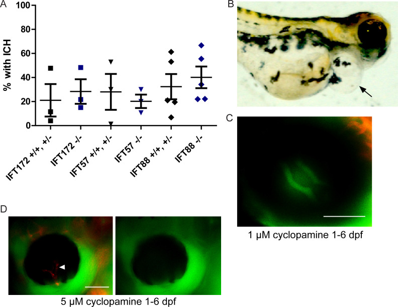
Fig 4
Zebrafish embryos were treated with 1 μM cyclopamine starting at 25 hpf. (A) Percentages of cyclopamine-treated IFT mutant and non-mutant embryos that developed ICH by 52 hpf. No significant differences were detected between mutants and non-mutants by Fisher’s exact test (p > 0.05). (B) Image of a 6 dpf wild-type fish that had cyclopamine-induced pericardial edema (arrow). (C) Fluorescent micrograph of the eye of a zebrafish treated with 1 μM cyclopamine from 1 to 6 dpf, demonstrating an intact blood-retinal barrier. (D) Fluorescent micrograph of a 5 μM cyclopamine-treated zebrafish at 6 dpf with non-perfusion of the hyaloid vasculature (arrowhead). Scale bars = 50 μm.