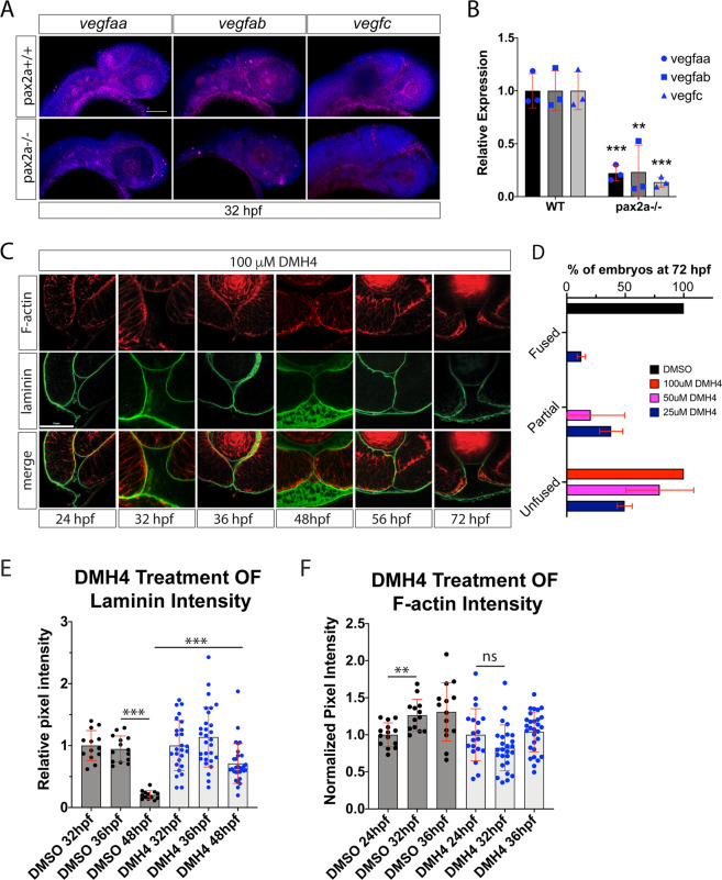
Inhibiting angiogenesis disrupts optic fissure fusion mechanics. (A) Fluorescent whole mount in situ hybridization of vegfaa, vegfab and vegfc at 32 hpf in WT and pax2a−/− embryos. Loss of pax2a results in decreased vegfaa, ab and c expression. DAPI is depicted in blue. Scale bar = 100 μm. (B) qPCR results for vegfaa, vegfab and vegfc expression from heads of pax2a+/+ and pax2a−/− embryos at 32 hpf. (C) 3D confocal images of Tg[kdrl:mCherry] (red) embryos treated with 100 μM DMH4 between 24-72 hpf. DNA was stained with DAPI (blue). Scale bar = 50 μm. (D) Quantification of 48 hpf DMH4 treated embryos for fissure fusion (absence of laminin signal), partial fusion (partial retention of laminin) or failure to fuse (retention of laminin throughout the fissure). n = 22 (DMSO), 52 (100 μM), 22 (50 μM), 24 (25 μM) ANOVA p < 0.0001. (E) Quantification of laminin signal intensity within the central-proximal region of the OF, normalized to regions of laminin staining juxtaposed to the lens. Relative pixel intensities are displayed. ANOVA p < 0.0001. (F) Quantification of F-actin signal intensity (phalloidin staining) within the central-proximal region of the OF, normalized to regions of F-actin signal within the lobe of the retina. Relative pixel intensities are displayed. ANOVA p < 0.0001.
This image is the copyrighted work of the attributed author or publisher, and
ZFIN has permission only to display this image to its users.
Additional permissions should be obtained from the applicable author or publisher of the image.
Full text @ Sci. Rep.
Your Input Welcome
Thank you for submitting comments. Your input has been emailed to ZFIN curators who may contact you if
additional information is required.
Oops. Something went wrong. Please try again later.