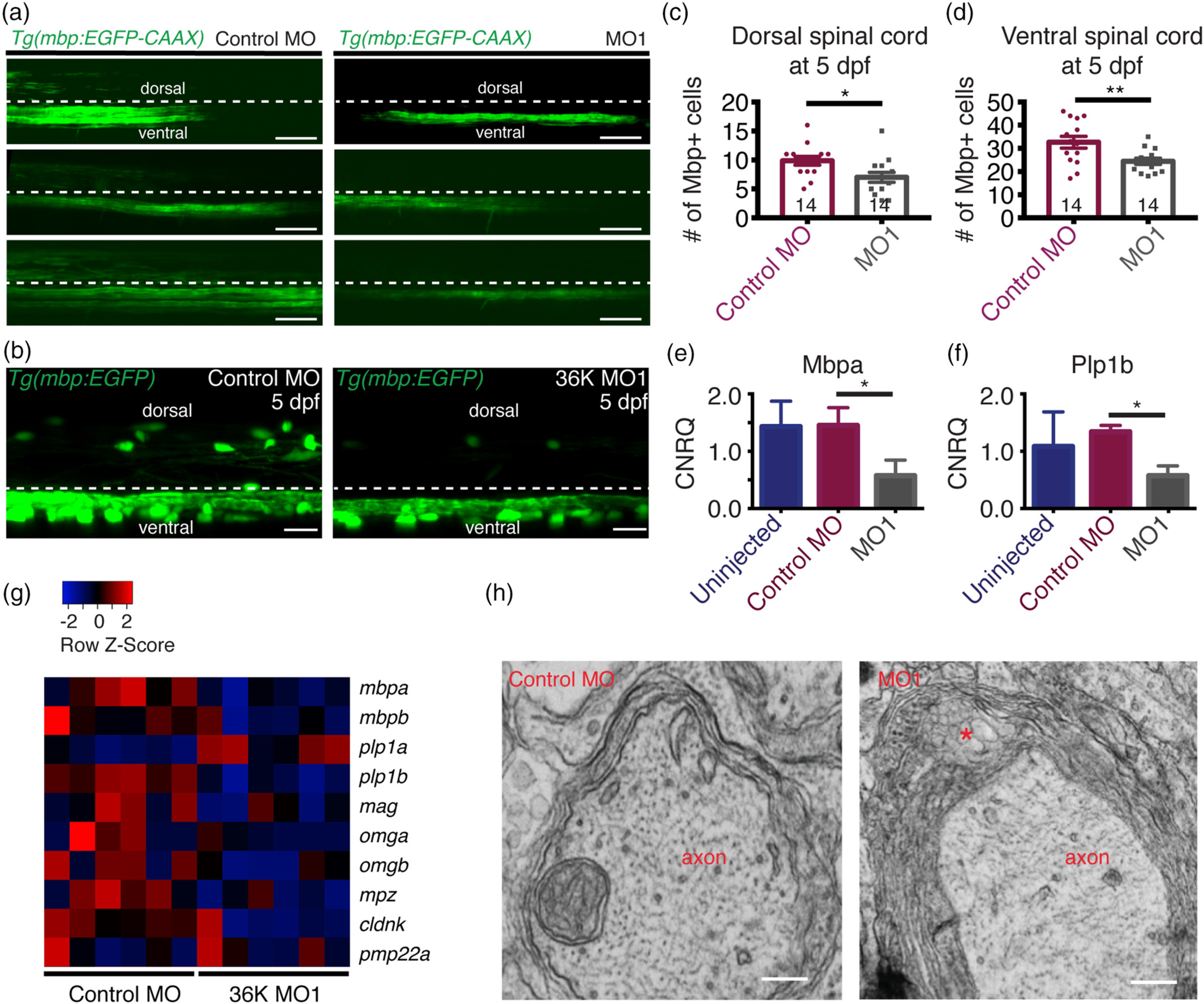
Fig. 4 36K knockdown zfl have less and disrupted myelin. (a) Representative images depicting lateral view of the spinal cord at 4 dpf of Tg(mbp:EGFP‐caax) fish injected with control MO and MO1 showing dorsal and ventral spinal cord regions separated by a dotted line, with the region above this line being dorsal and the region below the line the ventral spinal cord. Scale bar: 40 μm. (b) Representative image depicting lateral view of two spinal segments above the yolk extension at 5 dpf in Tg(mbp:EGFP) zfl injected with control MO and MO1. Scale bars: 20 μm. (c,d) Number of mbp positive cells in two spinal segments were significantly fewer in both the (c) dorsal spinal cord (d) and in the ventral spinal cord. Unpaired two tailed t test p‐value 0.0182 (*) for (c). Unpaired two tailed t test p‐value .0087 (**) for (d). (e,f) q‐rt‐PCR data showing calibrated normalized relative quantities (CNRQ) for (e) mbpa and (f) plp1b. Target genes were normalized to ß‐actin and ef1a. N = 5. Kruskal–Wallis ANOVA p ≤ .05 (*) followed by Dunn's multiple comparisons test for control MO versus MO1. (g) Heatmap generated from microarray analysis. Color‐coded data (red = upregulation, blue = downregulation) showing expression levels of different myelin genes in rows and different batches of 3 dpf larvae in columns. (h) Transmission electron micrographs showing myelin sheets wrapped around axons in control MO and MO1 zfl at 5 dpf. Asterisk represents noticeable cytoplasmic vacuolated structures in between myelin lamellae in MO1 larva. Scale bars: 0.3 μm
Image
Figure Caption
Figure Data
Acknowledgments
This image is the copyrighted work of the attributed author or publisher, and
ZFIN has permission only to display this image to its users.
Additional permissions should be obtained from the applicable author or publisher of the image.
Full text @ Glia

