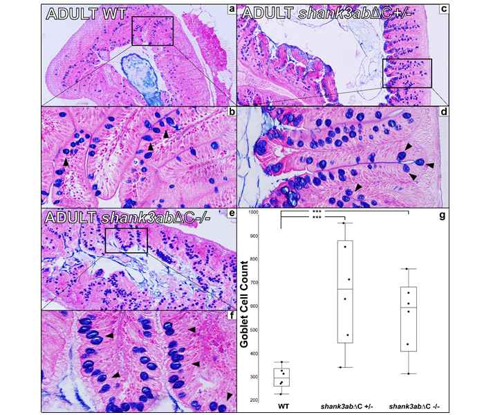
Fig. S2 Figure S2 a Transverse 5μm section of adult WT upper intestinal tissue stained with alcian blue and Eosin B (n=6 for WT, n=6 for shank3abΔC +/- and n=6 for shank3abΔC -/-). b 40x magnification shows dense plicae that extend to the point of nearly occluding the luminal space. c,d In shank3abΔC +/- upper intestinal tissue, increased counts of goblet cells (black arrowheads) suggest inflammation. e,f Homozygous shank3abΔC -/- adults also show increased goblet cell count. Comparison of goblet cell counts in WT, shank3abΔC +/-, and shank3abΔC -/- adults show a significant increase in the number of goblet cells in both shank3abΔC +/- and shank3abΔC -/- adults. WT and shank3abΔC -/- comparison (p<0.0138) and WT and shank3abΔC +/- comparison (p<0.0015).
Image
Figure Caption
Figure Data
Acknowledgments
This image is the copyrighted work of the attributed author or publisher, and
ZFIN has permission only to display this image to its users.
Additional permissions should be obtained from the applicable author or publisher of the image.
Full text @ Mol Autism

