Fig. 1
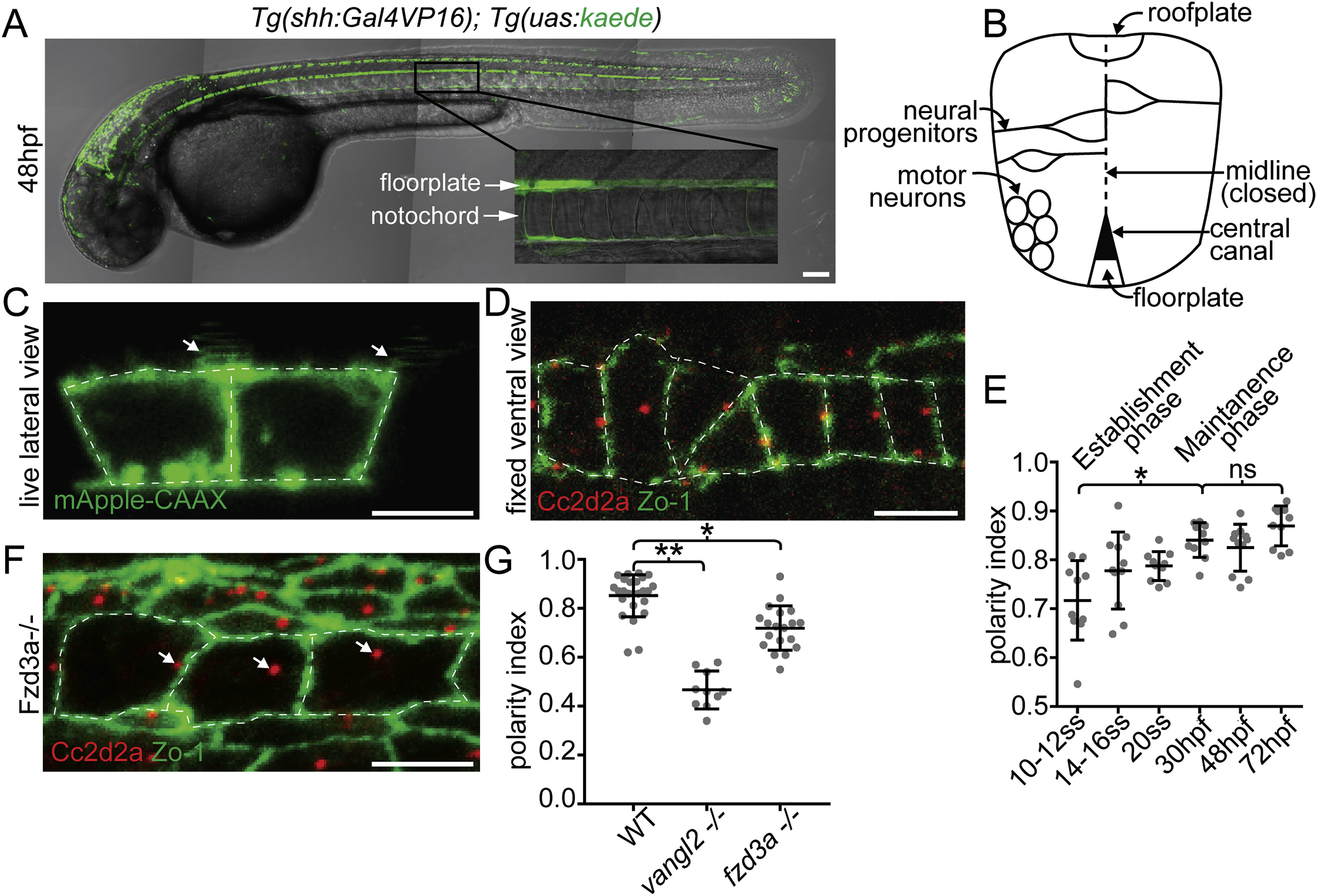
The floorplate is progressively planar polarized in a Vangl2 and Fzd3a-dependent manner. (A) A 48hpf Tg:(shh:gal4); Tg(uas:Kaede)zebrafish embryo expressing Kaede in the floorplate of the neural tube. (B) Schematic of a cross-section of the zebrafish neural tube at 48hpf (not to scale). (C) Single time point from a time lapse of a Tg(shh:gal4); Tg(uas:mApple-CAAX) embryo at 48hpf in which two adjacent floorplate cells are expressing membrane-localized mApple-CAAX (green). Posteriorly localized primary cilia (arrows) appear as squiggles due to their rapid motion. (D) Fixed ventral view of a 48hpf WT floorplate co-immunostained with ZO-1 to mark sub-apical tight junctions (green) and Cc2d2a to mark BBs (red). (E) Quantitation of per embryo polarity index from the 10–12ss through 72hpf. Each dot represents the average polarity index of at least 10 cells within a single embryo. Total N = 62 embryos, 1130 cells. *p < 0.0001; significance was determined with a Kruskal-Wallis test with Dunn's multiple comparison. (F) Fixed ventral view of a 48hpf fzd3a−/− floorplate stained as in D. White dotted line indicates position of cell boundaries, as determined by mApple-CAAX fluorescence (C) or ZO-1 staining (D,F), arrows indicate BB positions. (G) BB polarity indices of WT, vangl2−/− and fzd3a−/− embryos at 48hpf. Each dot represents the average polarity index of at least 10 cells within a single embryo. Total N = 54 embryos, 448 cells; **p < 0.0001; *p = 0.0018; significance was determined with a Kruskal-Wallis test with Dunn's multiple comparison. Anterior is to left in all images. Scale bars: 100 μm (A) or 5 μm (C,D,F).
Reprinted from Developmental Biology, 452(1), Mathewson, A.W., Berman, D., Moens, C.B., Microtubules are required for the maintenance of planar cell polarity in monociliated floorplate cells, 21-33, Copyright (2019) with permission from Elsevier. Full text @ Dev. Biol.

