
|
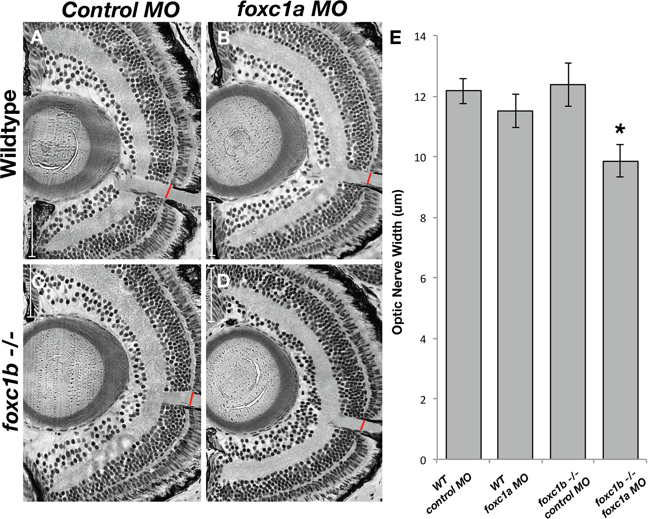
Fig. 3 Loss of foxc1 function reduced optic nerve thickness. foxc1a morpholino injection into a foxc1b -/- background reduced optic nerve thickness (D) when compared to wildtype embryos injected with standard negative control morpholinos (A), wildtype embryos injected with foxc1a morpholinos (B), foxc1b mutants injected with control morpholinos (C) This is statistically significant (E, comparison of A–D, p = 0.03 (C). Data presented as mean ± SEM. Red line indicates area of measurement. (For interpretation of the references to colour in this figure legend, the reader is referred to the web version of this article.)
Reprinted from Vision Research, 156, Umali, J., Hawkey-Noble, A., French, C.R., Loss of foxc1 in zebrafish reduces optic nerve size and cell number in the ganglion cell layer, 66-72, Copyright (2019) with permission from Elsevier. Full text @ Vision Res.