IMAGE

Fig. 3.

ID
ZDB-IMAGE-190723-918
Genes
Source
Figures for Gjini et al., 2019
Image
Figure Caption

Fig. 3.

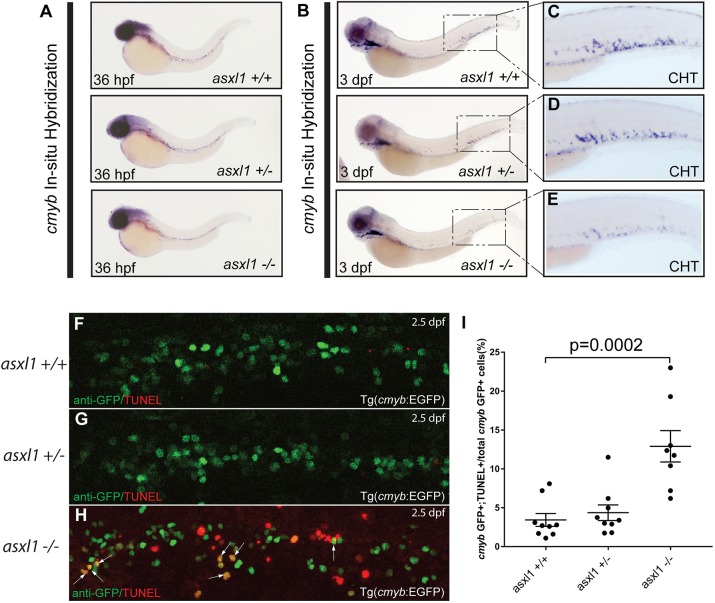
Loss of asxl1 induces apoptosis in cmyb-expressing embryonic HSPCs. WISH for cmyb was performed at 36 hpf (A) and 3 dpf (B-E) in asxl1+/+, asxl1+/ and asxl1−/− zebrafish embryos. Boxes in panel B are shown at higher magnification in C-E. (F-H) HSPCs (GFP; green) and cells undergoing apoptosis (TUNEL, TMR red, Roche) in the CHT of 48-hpf asxl1+/+, asxl1+/ and asxl1−/− zebrafish embryos in the Tg(cmyb:EGFP) reporter line background were identified by immunofluorescence microscopy following a dual TUNEL/anti-GFP assay. Arrows indicate apoptotic HSPCs with combined EGFP and DMR red fluorescence signals. (I) cmyb:GFP+/TUNEL+ cells in asxl1+/+, asxl1+/− and asxl1−/− were quantified as a percentage of total cmyb:GFP+ cells. Bars represent the mean and s.e.m. for 8-9 embryos each. Unpaired Student’s t-tests and Prism software were used to determine the P-value for each genotypic group compared with control cells.

Figure Data
Acknowledgments
This image is the copyrighted work of the attributed author or publisher, and ZFIN has permission only to display this image to its users. Additional permissions should be obtained from the applicable author or publisher of the image. Full text @ Dis. Model. Mech.