
|
Fig. 1
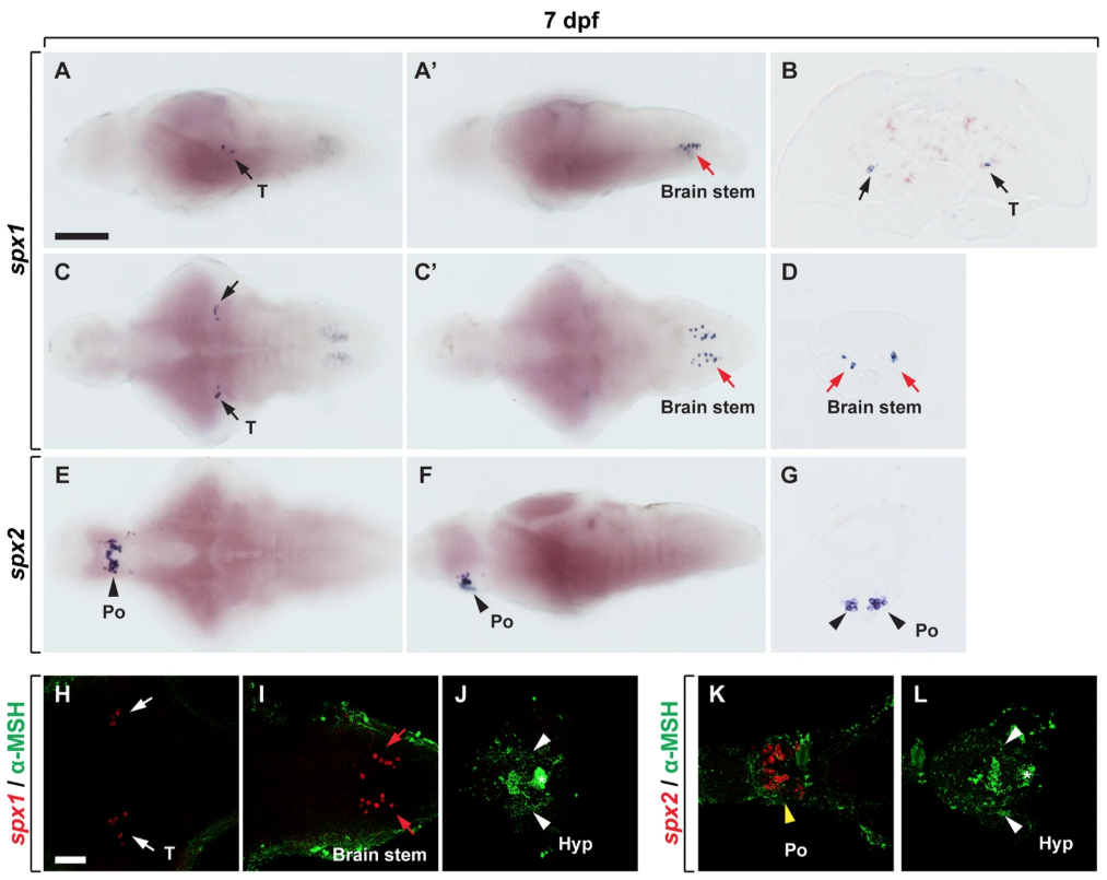
Expression of spx 1 and spx 2 mRNA in the brain of zebrafish larvae. (A–G) In situ RNA hybridization with spx1 (A–D) and spx2RNA probes (E–G) in the brain of zebrafish larvae at 7 days post-fertilisation (dpf). Lateral (A,A’,F), dorsal (C,C’), and ventral views (E) of the brain, anterior to the left. Transverse sections of the brain (B,D,G), dorsal to the top. (A–D) spx1 expression in the midbrain (A–C) and hindbrain (A’,C’,D). Black and red arrows indicate spx1 expression in the midbrain tegmentum and hindbrain, respectively. (E–G) spx2 mRNA expression in the preoptic area of the hypothalamus. Arrowheads indicate spx2expressing cells. (E–G,K,L). (H–L) Labelling with anti-α-melanocyte stimulating hormone (α-MSH) antibody following in situ RNA hybridization with spx1 (H–J) and spx2 (K,L). Ventral views with anterior to the left. White and red arrows indicate spx1-expressing cells in the midbrain (H) and hindbrain (I), and yellow arrowhead indicates spx2-expressing cells in the preoptic area (K). White arrowheads and asterisk label the hypothalamus and pituitary gland, respectively. Abbreviation: Hyp, hypothalamus; Po, preoptic region; T, midbrain tegmentum. Scale bar: 100 μm in A,A’,C,C’,E,F,E; 50 μm in B,D,G–L.