Fig. 6
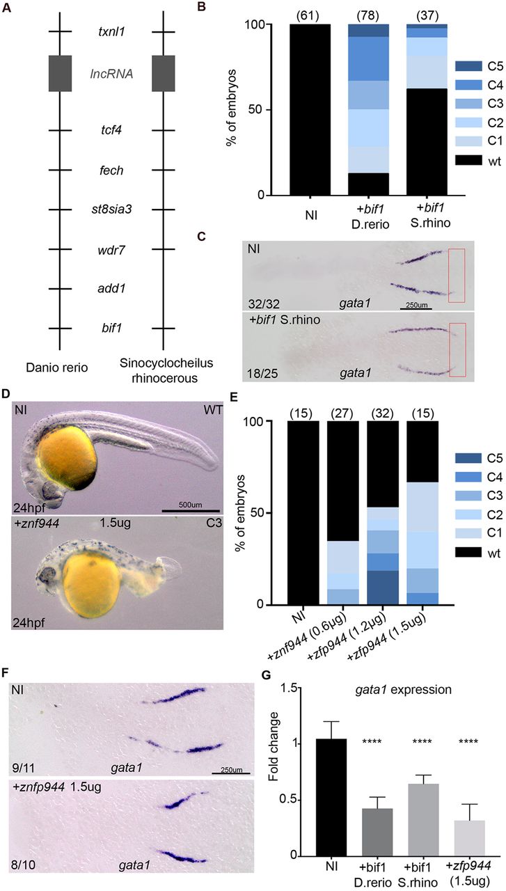
bif1 has orthologs in Sinocyclocheilus rhinocerous and in Mus musculus. (A) Synteny representation of the zebrafish and Sinocyclocheilus rhinocerous genome near to bif1. (B) Percentage of uninjected embryos and embryos injected with D. rerio or S. rhino bif1 mRNA showing a dorsalization morphology at 24 hpf in zebrafish embryos. (C) Whole-mount in situhybridization for gata1 at 12 hpf in non-injected embryos or embryos injected with bif1 S. rhinomRNA. Red rectangle indicates the region in which gata1a is ectopically expressed in bif1-overexpressing embryos. (D) Bright-field images of non-injected embryos or embryos injected with zfp944 mRNA at 24 hpf. (E) Percentage of uninjected and zfp944-injected zebrafish embryos showing a dorsalization morphology at 24 hpf. (F) Whole-mount in situ hybridization for gata1 at 12 hpf in non-injected embryos or embryos injected with zfp944 mRNA. (G) Quantification of gata1 by qPCR, in 48 hpf embryos, either non-injected or injected with D. rerio bif1, S. rhino bif1 or murine Zfp944 mRNA (n=3 biological replicates for all). Statistical analysis was carried out using an unpaired Student t-test. Data are mean±s.e.m. ****P<0.0001.

