Fig. 1
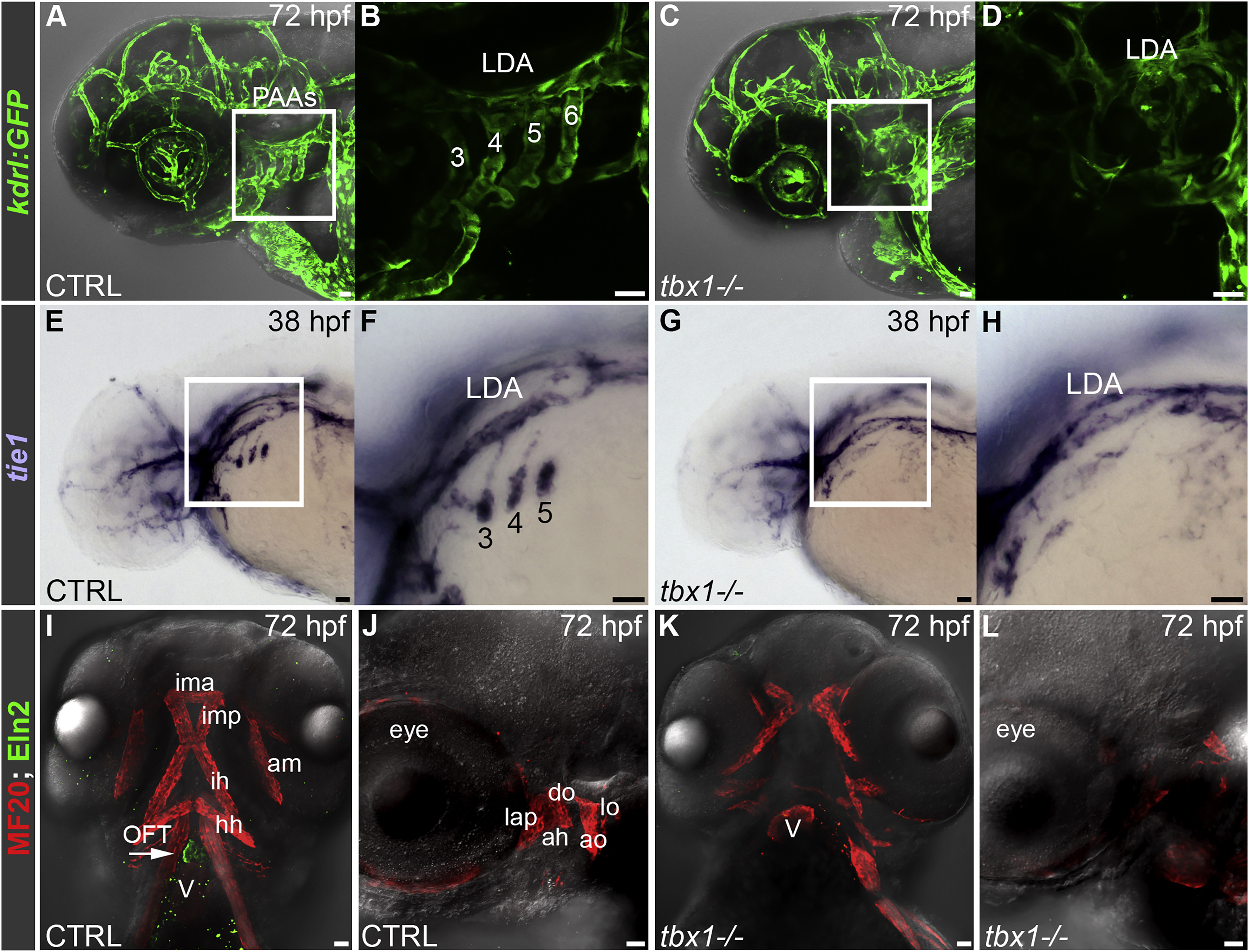
Tbx1 Is Required for Pharyngeal Arch Artery, Head Muscle, and Outflow Tract Morphogenesis in Zebrafish
(A–D) Confocal z-stacks of the head (A and C) and pharyngeal arch arteries (PAAs; B and D) in live 72 hr post-fertilization (hpf) control (CTRL; A and B; n = 60) and tbx1 mutant (C and D; n = 20) embryos carrying the Tg(kdrl:GFP) endothelial reporter. (B) and (D) are independent z-stacks acquired at higher magnification of the boxed regions in (A) and (C). Merged bright-field images and confocal z-stacks are shown in (A) and (C). The numbers in (B) identify each PAA. Lateral views, anterior left.
(E–H) Bright-field z-stacks of the head (E and G) and pharyngeal arches (F and H) in 38 hpf CTRL (E and F; n = 40) and tbx1 mutant (G and H; n = 19) embryos processed by in situ hybridization with a tie1 riboprobe. The boxed regions in (E) and (G) are shown at higher magnification in (F) and (H). The numbers in (F) identify each tie1+ PAA angioblast cluster. Lateral views, anterior left.
(I–L) Merged bright-field images and confocal z-stacks of the head region in 72 hpf CTRL (I and J; n = 40) and tbx1 mutant (K and L; n = 19) embryos co-immunostained with antibodies recognizing striated muscle (MF20, red) or OFT smooth muscle (Eln2, green). Ventral views, anterior up in (I) and (K). Lateral views, anterior left in (J) and (L). Arrow in (I) highlights the Eln2+ OFT smooth muscle that is missing in tbx1 mutants. In all cases, little to no variation was observed between animals in each experimental group.
Scale bars, 25 μm. Pharyngeal arch1 (PA1)-derived head muscles: am, abductor mandibulae; do, dilator operculi; ima, intermandibularis anterior; imp, intermandibularis posterior; lap, levator arcus palatini; PA2-derived head muscles: ah, adductor hyoideus; ao, adductor operculi; hh, hyohyoidus; ih, interhyoidus; lo, levator operculi. LDA, lateral dorsal aorta; OFT, outflow tract; PAAs, pharyngeal arch arteries; V, ventricle.

