Image
Figure Caption
Fig. 5
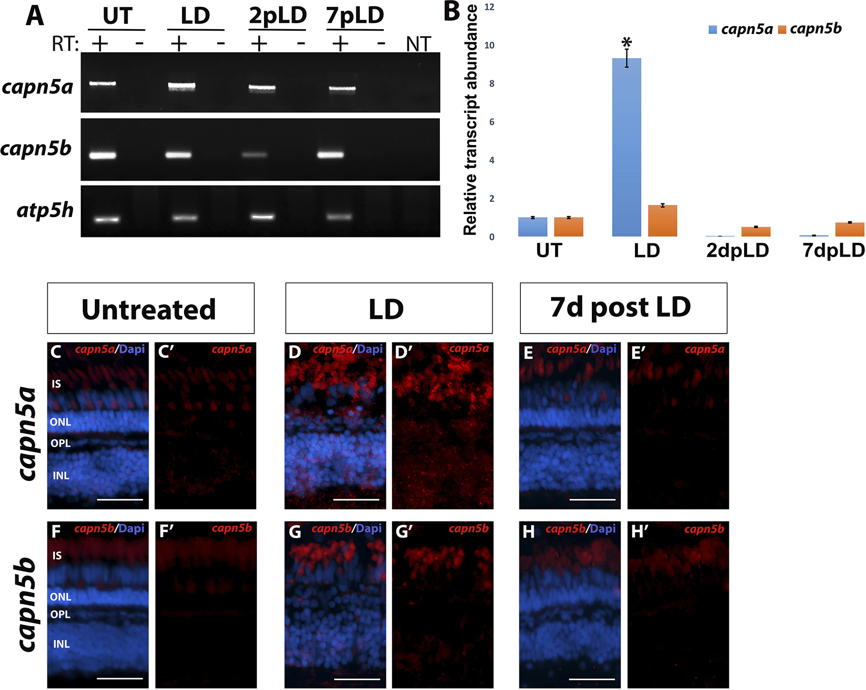
Capn5a expression is induced in response to acute LD. (A) RT-PCR of capn5a and capn5b expression during and following acute LD. A significant increase in expression of capn5a was observed after 3 days of LD, which returned to WT levels by 2 days post LD. Capn5b expression did not change during LD. (B) qPCR for capn5a and capn5b during and following LD. A 12-fold increase in capn5a expression was observed in the LD retina compared with untreated retina, followed by a significant decrease in expression post LD. Capn5b expression did not change during or after LD. (C–E′) FISH for capn5a in untreated retina (UT), during LD, and post LD (7 days post LD). Whereas capn5a expression in the UT retina was confined to the OPL and cone IS, capn5a expression in the LD retina was seen in the INL as well as the OPL and IS. (F–H′) FISH for capn5b in the UT, LD, and 7 days post LD retina. The capn5b expression pattern did not change during or after LD. Scale bars: 50 μm. *P < 0.05.
Figure Data
Acknowledgments
This image is the copyrighted work of the attributed author or publisher, and
ZFIN has permission only to display this image to its users.
Additional permissions should be obtained from the applicable author or publisher of the image.
Full text @ Invest. Ophthalmol. Vis. Sci.

