Fig. 2
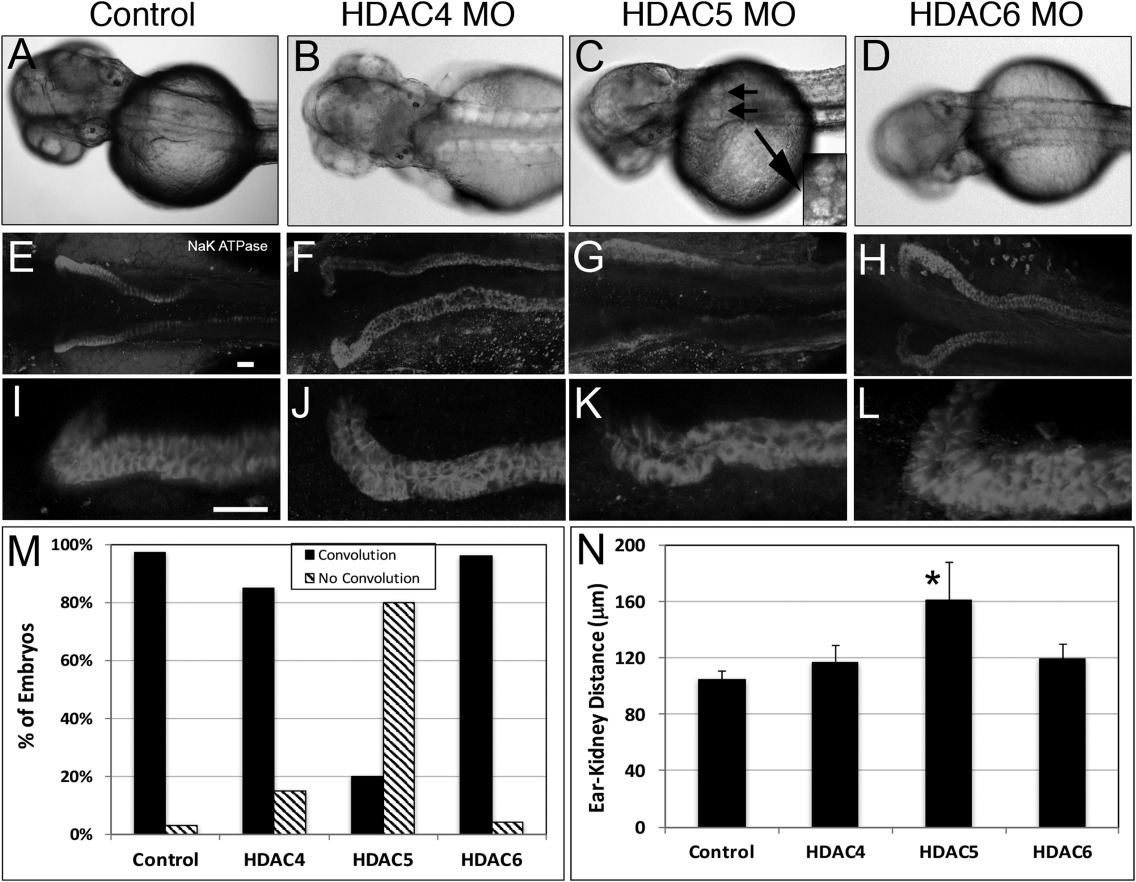
Kidney cysts form in hdac5 morphants. A–D: Dorsal images of embryos injected with 2 ng control, 2 ng HDAC4, 2 ng HDAC5, or 8 ng HDAC6 MO. Cysts were visible in hdac5 morphants (small arrows and inset, magnified view) at 48 hpf. Morphant embryos at 72 hpf were immunostained for α1 Na+K+ ATPase and imaged at low magnification (E–H) and high magnification (I–L). Scale bar = 100 μm. Anterior migration (convolution) is blocked in hdac5 morphants as summarized across the population by image analysis (M) and by the increase in distance between the anterior kidney and posterior ear (N). n = 40–70 embryos per experiment. *P < 0.005.

