
|
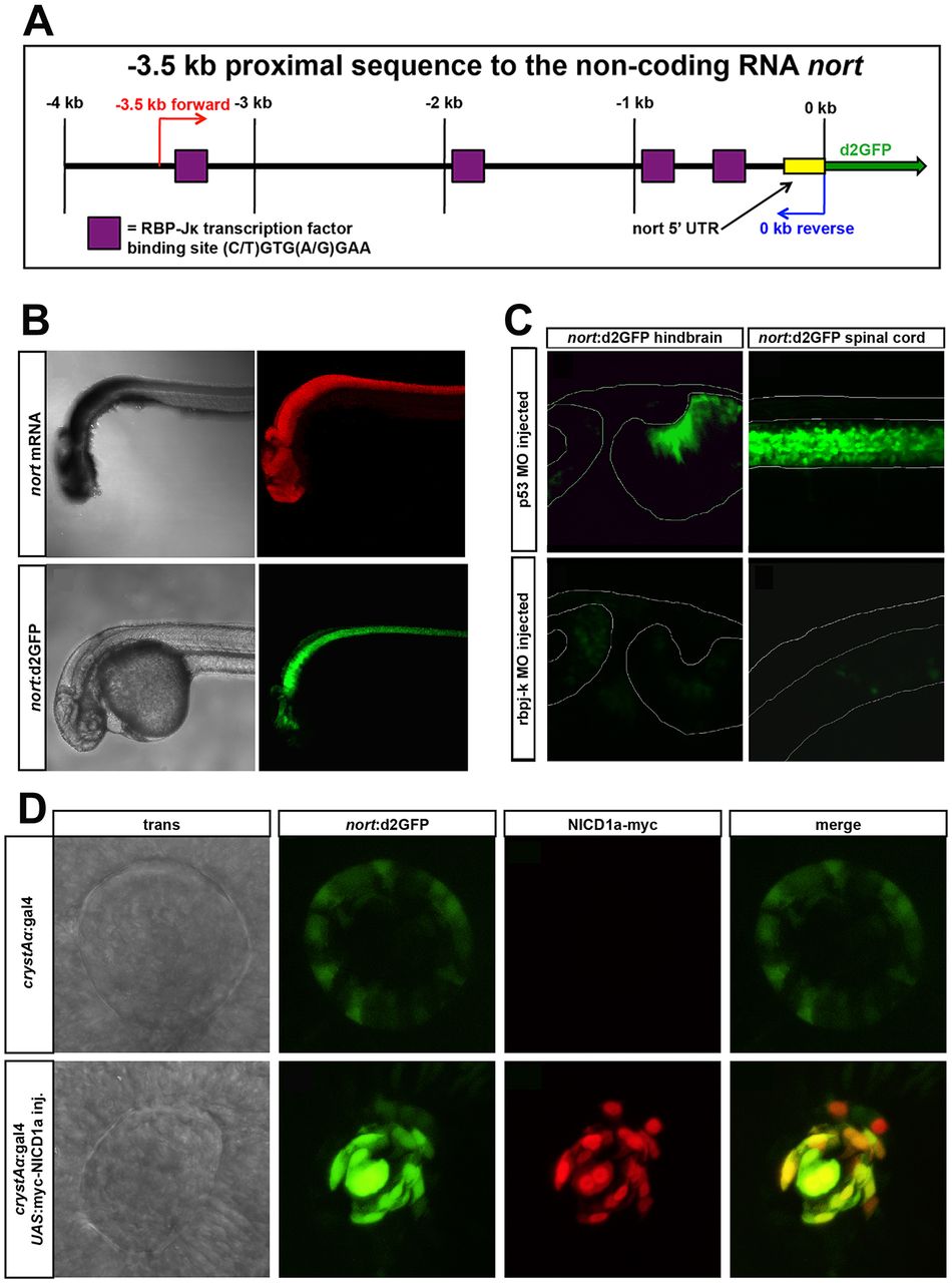
Fig. 4
A nort transgenic reporter for Notch activity. (A) Schematic of the 3.5 kb nort proximal promoter. This region contains four canonical rbpj-K-binding sites. (B) nort mRNA (red) and nort:d2GFP (green) transgene expressed in the same tissues at 30 hpf. (C) nort:d2GFP expression was reduced in a rbpj-k MO-injected 48 hpf hindbrain and spinal cord compared with the p53 MO-injected controls. The morphology of the hindbrain was similar to the controls, whereas the rbpj-k MO-injected spinal cord was curved. (D) Normal expression of nort:d2GFP is found in the lens epithelium. Overexpression of myc-NICD1a in the lens using crystAα:gal4 causes enhanced ectopic nort:d2GFP expression. Myc immunofluorescence colocalizes with upregulated d2GFP at 28 hpf.