
|
Fig. S7
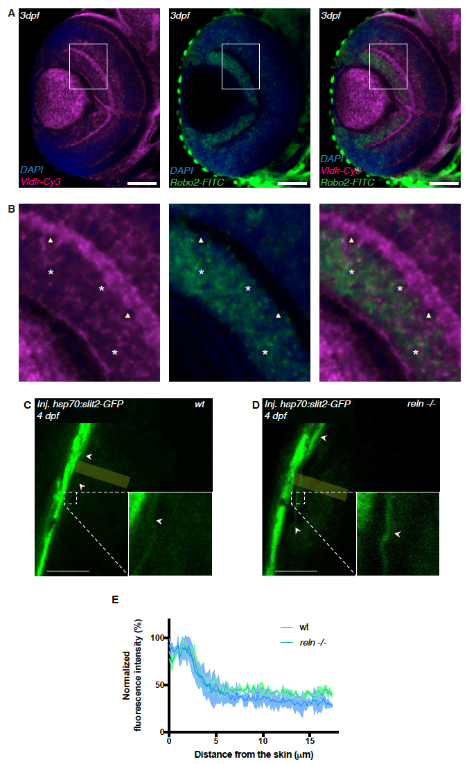
vldlr and robo2 expression in RGCs of the developing retina and Slit distribution in the tectum. Related to Figure 7.
(A) Confocal cross-section of a 3 dpf zebrafish retina showing mRNA expression of the Reelin receptor vldlr (magenta) and robo2 (green) in RGCs. Scale bars = 50 μm.
(B) Insets as indicated in (A) showing mostly an overlap of vldlr and robo2 expression in RGCs (white asterisk) consistent with a model according to which RGCs integrate both Reelin and Slit signals to identify the single target lamina in the tectum. In a few RGCs we could only detect vldlr (white triangles) possibly owing to the in situ probe not being sensitive enough to pick up on low amounts of the robo2 mRNA.
(C) Confocal section through the tectum of a 4dpf wild-type larva injected with hsp70:slit2- GFP at one-cell stage and heat-shocked at 76 hpf. Slit2-GFP (green) is enriched at the surface of the tectal neuropil (arrows). The yellow rectangle indicates the dimension along which the slit signal was measured. Scale bar = 30μm
(D) Confocal section through the tectum of a 4dpf reln -/- larva injected with hsp70:slit2-GFP at one-cell stage and heat-shocked at 76 hpf. Slit2-GFP (green) is enriched at the surface of the tectal neuropil (arrows). The yellow rectangle indicates the dimension along which the slit signal was measured. Scale bar = 30μm
(E) Densitometric plots of normalized GFP fluorescence intensity in 4 dpf wild-type (along yellow rectangle in C) and reln -/- (along yellow rectangle in D) tecta. Slit2-GFP localization in the neuropil is indistinguishable between wild-type and reln -/- tecta.