Fig. S2
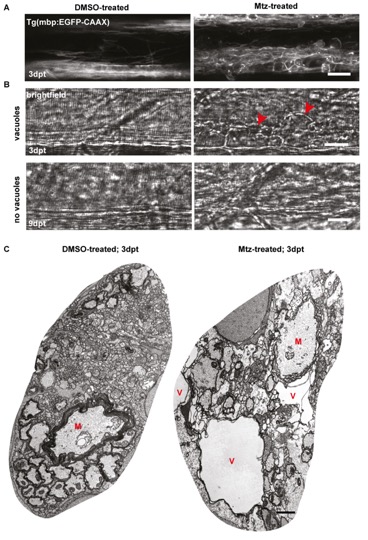
Oligodendrocyte ablation results in extensive myelin vacuolation.
A. Oligodendrocytes labelled with mbp:EGFP-CAAX in DMSO- or Mtz-treated Tg(mbp:mCherry-NTR) animals, before (top panel) or after (bottom panel) a two-day treatment with Mtz. Red arrowheads indicate putative myelin vacuoles.
A. Representative example of myelin vacuolation and disruption of spinal cord organisation, as visualised by the stable transgenic line Tg(mbp:EGFP-CAAX).
B. Brightfield images taken at 3dpt (top), where large vacuoles are clearly visible in the Mtz-treated animal (red arrowheads) and at 9dpt (bottom), when vacuoles are no longer detectible.
C. Representative electron micrographs of ventral hemi-spinal cords of DMSO- and Mtz-treated Tg(mbp:mCherry-NTR) animals. The control image shows the typical organisation of the spinal cord at this age, whereas the treated image contains numerous large fluid-filled vacuoles (labelled with red letters V) which disrupt the overall structure of the spinal cord.
Scale bars in A-B: 20μm.
Image
Figure Caption
Acknowledgments
This image is the copyrighted work of the attributed author or publisher, and
ZFIN has permission only to display this image to its users.
Additional permissions should be obtained from the applicable author or publisher of the image.
Full text @ PLoS One

