Fig. 5
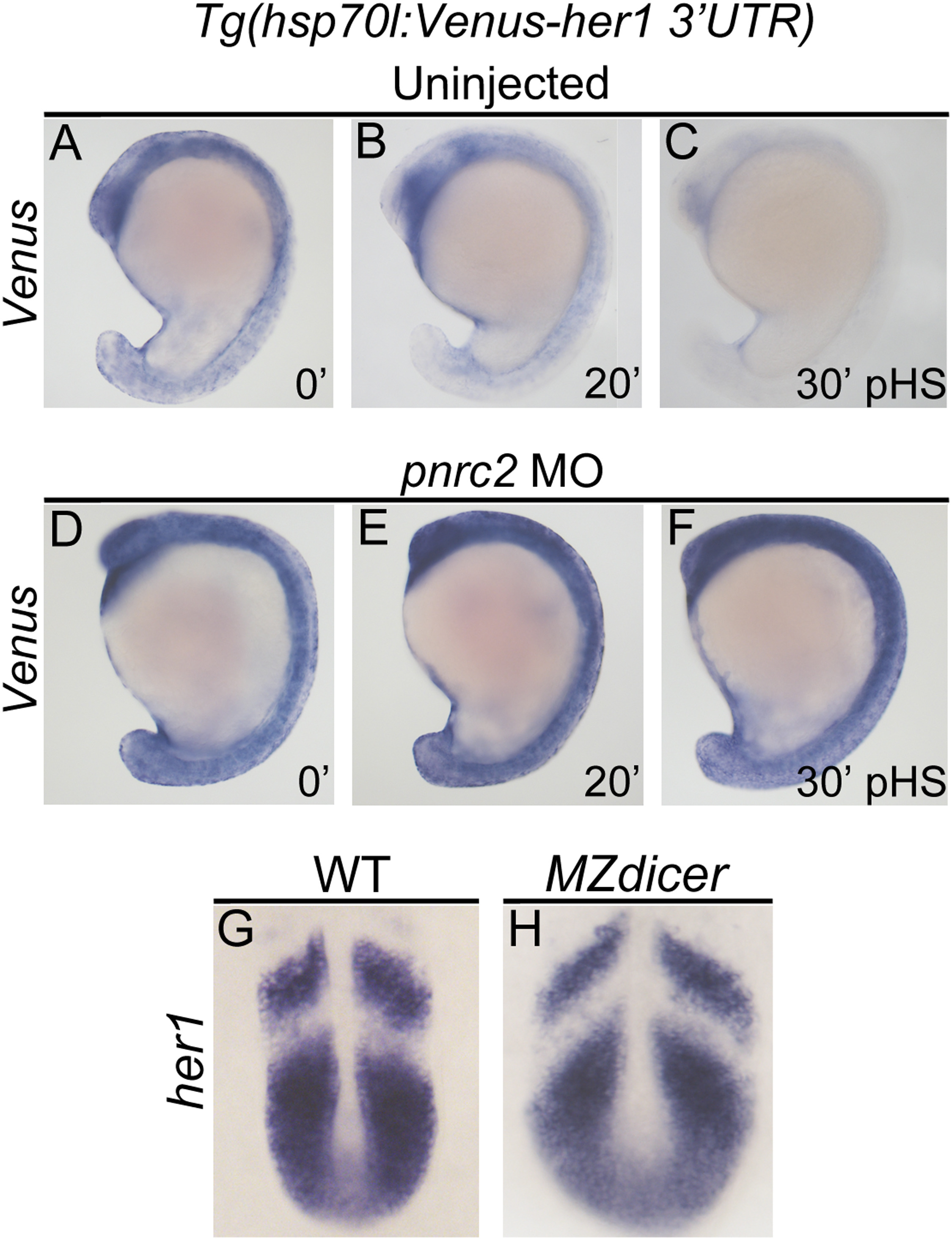
Pnrc2-mediated decay functions via 3’UTR recognition and does not require Dicer-dependent miRNAs. Stably transgenic embryos carrying the hsp70l:Venus her1 3’UTR reporter were injected at the 1-cell stage with pnrc2 sbMO or reserved as uninjected controls. Embryos were then raised to mid-segmentation stage, heat-shocked for 15 min, then collected at the indicated minutes post-heat-shock (pHS) and processed by Venus in situ hybridization (n=65, 6 ng pnrc2 sbMO; n=55, uninjected controls) (A-F). Venus transcripts are not detected in the absence of heat-shock (n=10) (not shown). To determine whether Dicer-generated miRNAs contribute to her1 mRNA decay, MZdicer mutants (n=11) and wild-type controls (n=10) were raised to mid-segmentation stages (16–18 hpf) and processed by her1 in situ hybridization (G, H). At this timepoint, MZdicer mutants are developmentally delayed relative to wild-type controls (Giraldez et al., 2005), and thus have a different overall shape.
Reprinted from Developmental Biology, 429(1), Gallagher, T.L., Tietz, K.T., Morrow, Z.T., McCammon, J.M., Goldrich, M.L., Derr, N.L., Amacher, S.L., Pnrc2 regulates 3'UTR-mediated decay of segmentation clock-associated transcripts during zebrafish segmentation, 225-239, Copyright (2017) with permission from Elsevier. Full text @ Dev. Biol.

