Image
Figure Caption
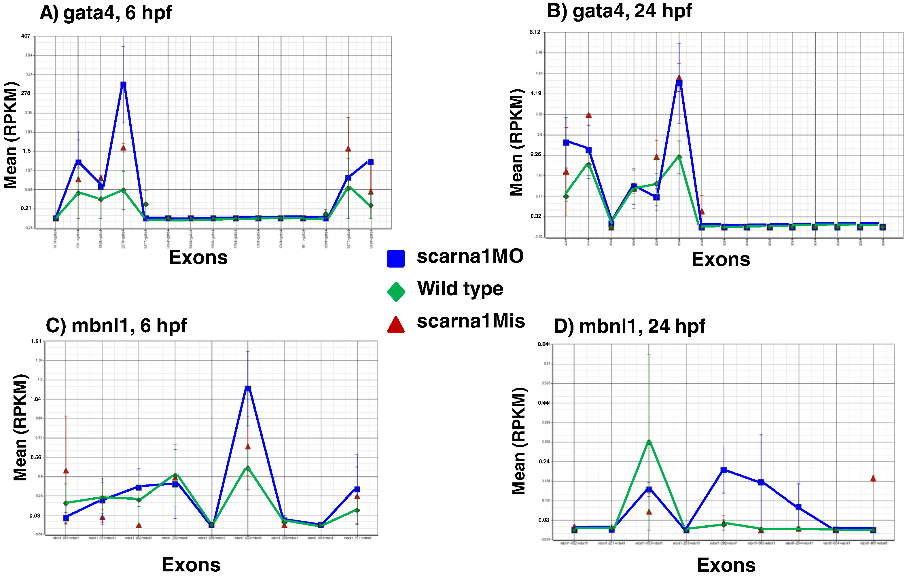
Fig. 7
Representative examples of splice isoform variation (exon retention) after treating zebrafish embryos with anti-scarna1 morpholino. Each point on the horizontal axis represents an exon and the value represents the mean RPKM (reads per kilobase per million) for that exon. Gata4 and Mbnl1, 6 and 24 h post fertilization (hpf).
Figure Data
Acknowledgments
This image is the copyrighted work of the attributed author or publisher, and
ZFIN has permission only to display this image to its users.
Additional permissions should be obtained from the applicable author or publisher of the image.
Reprinted from Biochimica et biophysica acta. Molecular basis of disease, 1852(8), Patil, P., Kibiryeva, N., Uechi, T., Marshall, J., O'Brien, J.E., Artman, M., Kenmochi, N., Bittel, D.C., scaRNAs Regulate Splicing and Vertebrate Heart Development, 1619-29, Copyright (2015) with permission from Elsevier. Full text @ BBA Molecular Basis of Disease

