Fig. S2
extl3/ext2 mutants show a late onset phenotype. Related to Figure 1-3.
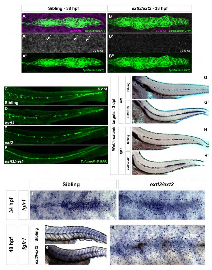
(A-B′′) extl3/ext2 mutants show significant downregulation of HS chains as evidenced by 3G10 antibody whole mount staining. (C-F) LL phenotypes in 5 dpf extl3/ext2 mutants. Compared to ext2 and extl3/ext2 mutants, extl3 mutants show only mild defects in the LL at this stage of development (D). ext2 mutant primordia do not reach the tail tip by 5 dpf and possess an elongated tip but clear neuromast formation (E). extl3/ext2 mutants are characterized by a collapsed LL where all neuromasts have lost their round shape, likely due to the loss of Fgf signaling (F). (G-H′) Wnt/β-catenin targets lef1 and fgf3 are expanded along the whole lateral line in 3 dpf extl3/ext2 mutants. (I-K′) The Fgf target fgfr1 is progressively downregulated between 34 hpf and 3 dpf in extl3/ext2 mutants. (K′) Close up of a 48 hpf extl3/ext2 primordium showing the downregulated fgfr1 expression.

