
Image
|
Figure Caption
Fig. S3
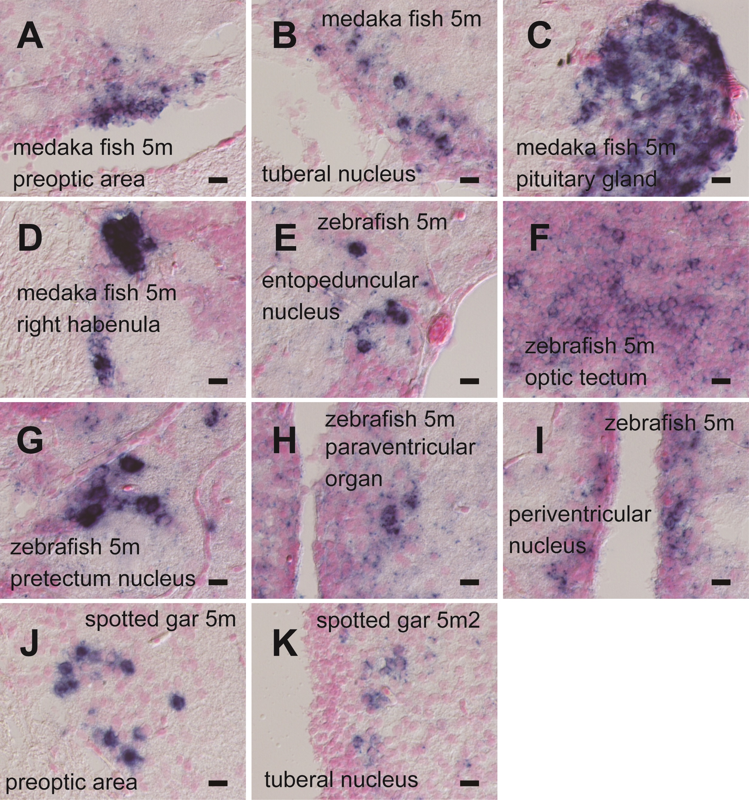
High-power field microscopic images of in situ hybridization in fish brains (600x magnification).
(A) Medaka fish Opn5m in preoptic area. (B) Medaka fish Opn5m in tuberal nucleus. (C) Medaka fish Opn5m in pituitary gland. (D) Medaka fish Opn5m in right habenula. (E) Zebrafish Opn5m in entopeduncular nucleus. (F) Zebrafish Opn5m in optic tectum. (G) Zebrafish Opn5m in pretectal nucleus. (H) Zebrafish Opn5m in paraventricular organ. (I) Zebrafish Opn5m in periventricular nucleus. (J) Spotted gar Opn5m in preoptic area. (K) Spotted gar Opn5m2 in tuberal nucleus. Scale bar: 10 µm.
Figure Data
Acknowledgments
This image is the copyrighted work of the attributed author or publisher, and
ZFIN has permission only to display this image to its users.
Additional permissions should be obtained from the applicable author or publisher of the image.
Full text @ PLoS One