Fig. 4
Fig. 4
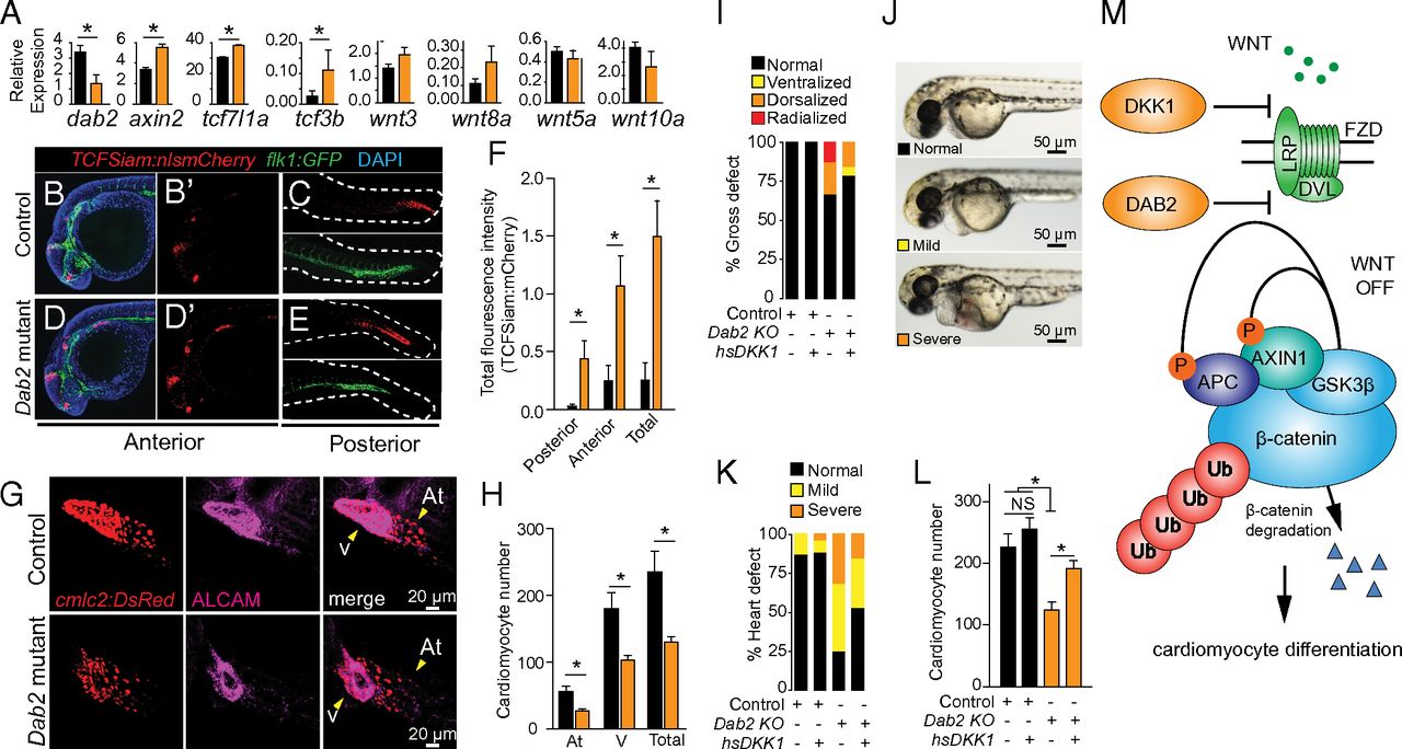
Dab2 promotes cardiomyocyte development by negatively regulating WNT/β-catenin signaling. (A) qRT-PCR of dab2 and a panel of known WNT/β-catenin target genes (axin2, tcf7I1a, and tcf3b) and ligands (wnt3, wnt8a, wnt5a, and wnt10a) in control (black bars) and Dab2 mutants (orange bars) at 24 hpf. (B–E) Representative brightest point projection confocal image (Z series) of control (B and C) and Dab2 mutants (D and E) at 24 hpf. TCFSiam:nlsmCherry is shown in red, flk1:GFP in green, and DAPI in blue. Panels shown are anterior merged (B and D), anterior red (B′ and D′), and posterior red and green (C and E). (F) Quantification of total fluorescence intensity between control and Dab2 mutants at 24 hpf. n ≥3 biological replicates. *P ≤ 0.05, Student’s t test. Data are mean ± SEM. (G) Representative brightest point projection confocal (Z series) images of control (Top) and Dab2 mutants (Bottom) at 48 hpf. cmlc2:DsRed-nuc is in red (Left), and ALCAM is in magenta (Middle) (H) Quantification of atrial (At) and ventricular (V) cardiomyocytes. (I) Percent gross morphological defect scoring in control and Dab2 mutants after overexpression of DKK1 scored at 48 hpf. (J) Representative images of normal, mild, and severe heart defects at 48 hpf. (K) Percent heart defects in control and Dab2 mutants after overexpression of DKK1 scored at 48 hpf. (L) Quantification of total cardiomyocyte number. *P ≤ 0.05, Student’s t test. n = 30–45 per group for heat shock and scoring for panels I and K. For quantification of cardiomyocyte nuclei, n = 4–6 fish hearts per condition analyzed by z-series confocal microscopy. Data are mean ± SEM. (M) Model: DAB2 inhibition of WNT/β-catenin signaling promotes cardiomyocyte differentiation.

