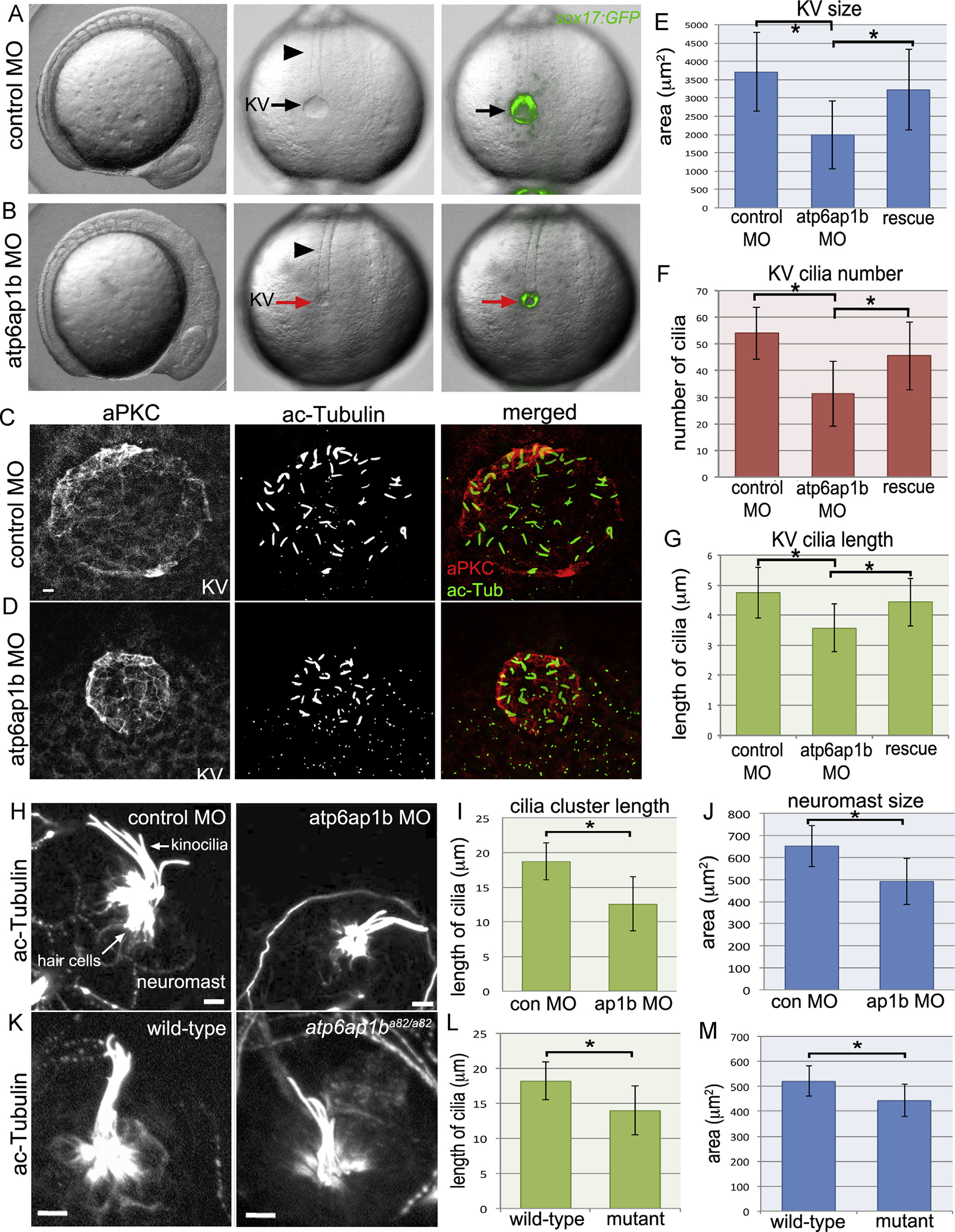
Fig. 2
Atp6ap1b depletion altered KV organ size and cilia formation. (A, B) Analysis of live Tg(sox17:GFP) embryos injected with control (A) or atp6ap1b MO (B) at the 8 somite stage. atp6ap1b MO embryos were indistinguishable from controls except for the small size of KV (arrows) labeled by GFP expression. The notochord (arrowheads) appeared normal in atp6ap1b MO embryos. (C, D) Immunofluorescent staining of KVs using aPKC antibodies to label apical membranes and acetylated tubulin (ac-Tub) antibodies to label cilia in control (C) or atp6ap1b (D) MO embryos. (E–G) Quantification of KV area (E), KV cilia number (F), and KV cilia length (G) in control MO (n=32) embryos, atp6ap1b MO injected (n=53) embryos and rescue (n=34) embryos co-injected with atp6ap1b MO+atp6ap1b mRNA. (H-M) Analysis of neuromasts stained with ac-Tub antibodies at 3 dpf in control MO and atp6ap1b MO embryos (H) or wild-type and atp6ap1ba82/a82 mutants (K). Kinocilia cluster length and neuromast area were reduced in atp6ap1b MO injected (n=13) embryos relative to controls (n=13) (I–J) and in atp6ap1ba82/a82 mutants (n=7) relative to wild-type (n=7) siblings (L–M). All scale bars=5 µm.
Reprinted from Developmental Biology, 407(1), Gokey, J.J., Dasgupta, A., Amack, J.D., The V-ATPase accessory protein Atp6ap1b mediates dorsal forerunner cell proliferation and left-right asymmetry in zebrafish, 115-30, Copyright (2015) with permission from Elsevier. Full text @ Dev. Biol.

