
|
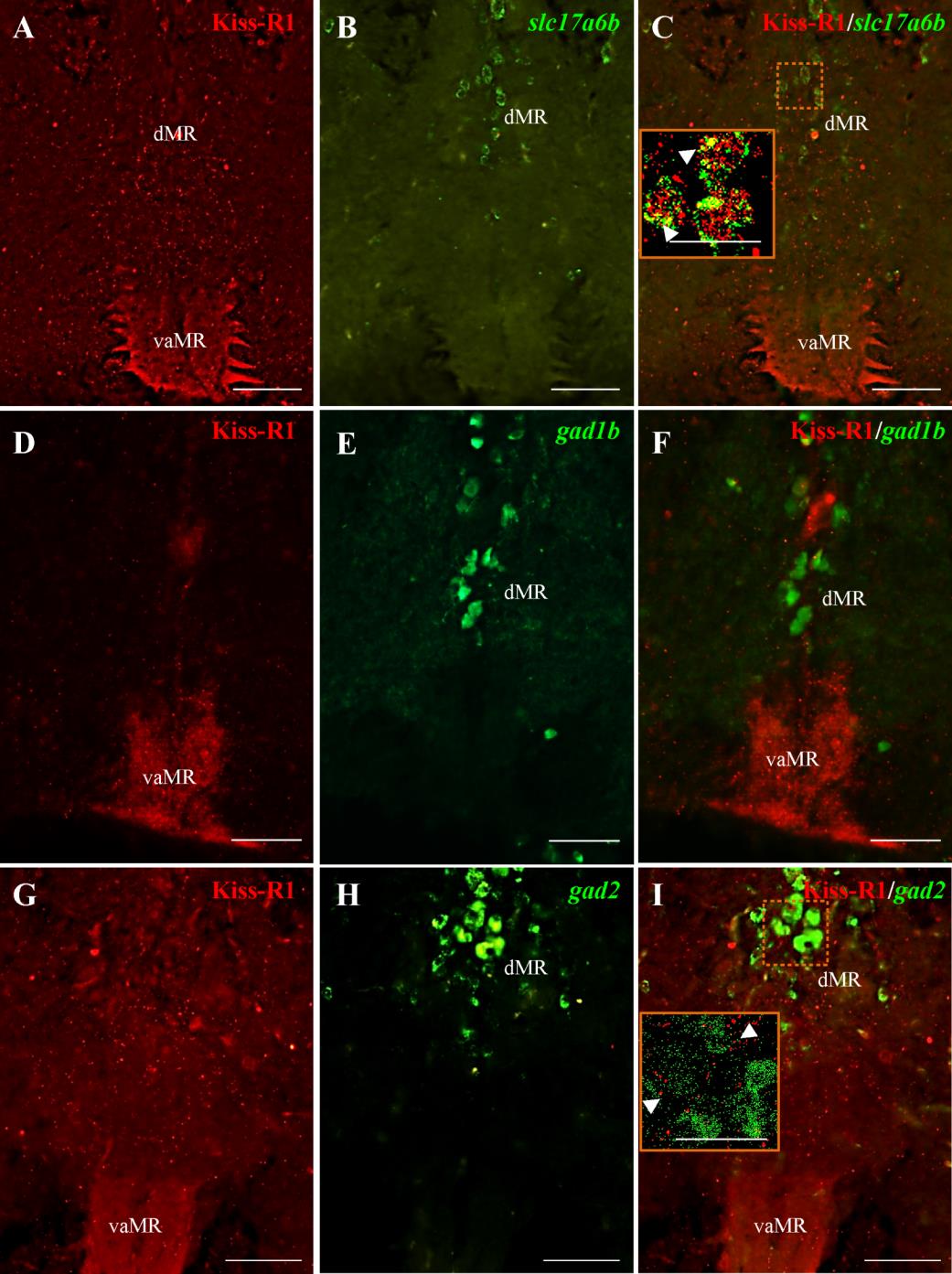
Fig. S3 Dual-fluorescence labeling of Kiss-R1 (red) in glutamatergic and GABAergic neurons (green) in raphe nuclei. A-C: Kiss-R1-immunoreactivity co-expressed with slc17a6b-expressing cells as denoted by the confocal image (inset C; 79.8X; N.A. = 1.4; z-step 0.15 µm). D-F: GFP-labeled Gad1b neurons were only observed in the dMR with no close associations observed with Kiss-R1 fibers. G-I: There was no co-localization of Kiss-R1 with gad2- expressing cells, but Kiss-R1-ir fibers were noted within close proximity in the dMR. Presence of actual space of at least 0.15 µm noted between fibers and cells (inset I). Scale bars, A-F: 100 µm and inset C and I: 50 µm.