
|
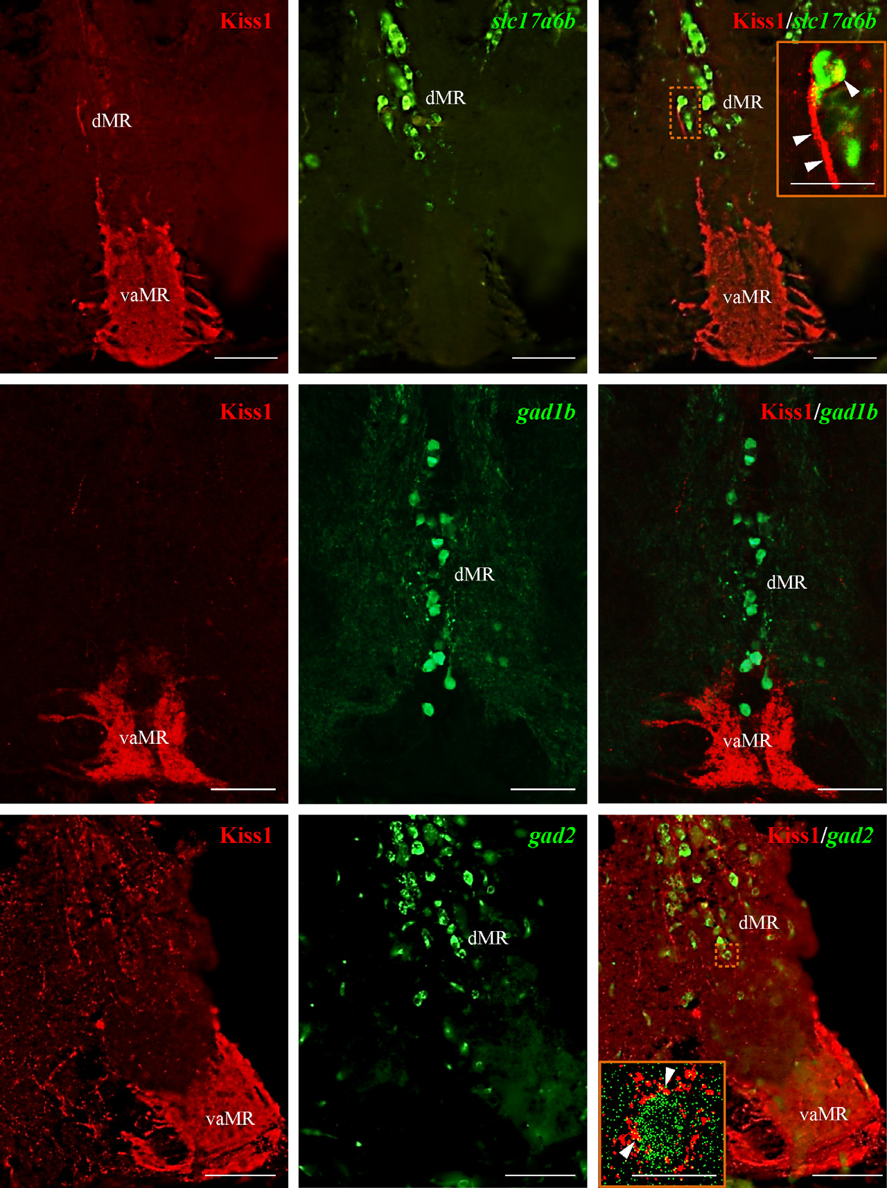
Fig. 7
Dual-fluorescence labeling of kisspeptin 1 (Kiss1) (red) in glutamatergic and GABAergic neurons (green) in raphe nuclei. (a–c) In the raphe nuclei, Kiss1-immunoreactive (-ir) fibers were seen in close association with slc17a6b mRNA expressing neurons in the dMR as denoted by the confocal image (inset C). (d–f) green fluorescent protein (GFP)-labeled Gad1b neurons were observed only in the dMR with no close associations observed with Kiss1-ir fibers. G-I: Kiss1-ir fibers were seen in close association with gad2 mRNA expressing neurons in the dMR, denoted by the confocal image (inset I; 60× plus 1.5× optical zoom; N.A. = 1.4; z-step = 0.15 µm). Presence of actual space of at least 0.15 µm noted between fibers and cells. Scale bars, (a–i): 100 µm; inset C and I: 50 µm.