
|
Fig. 6
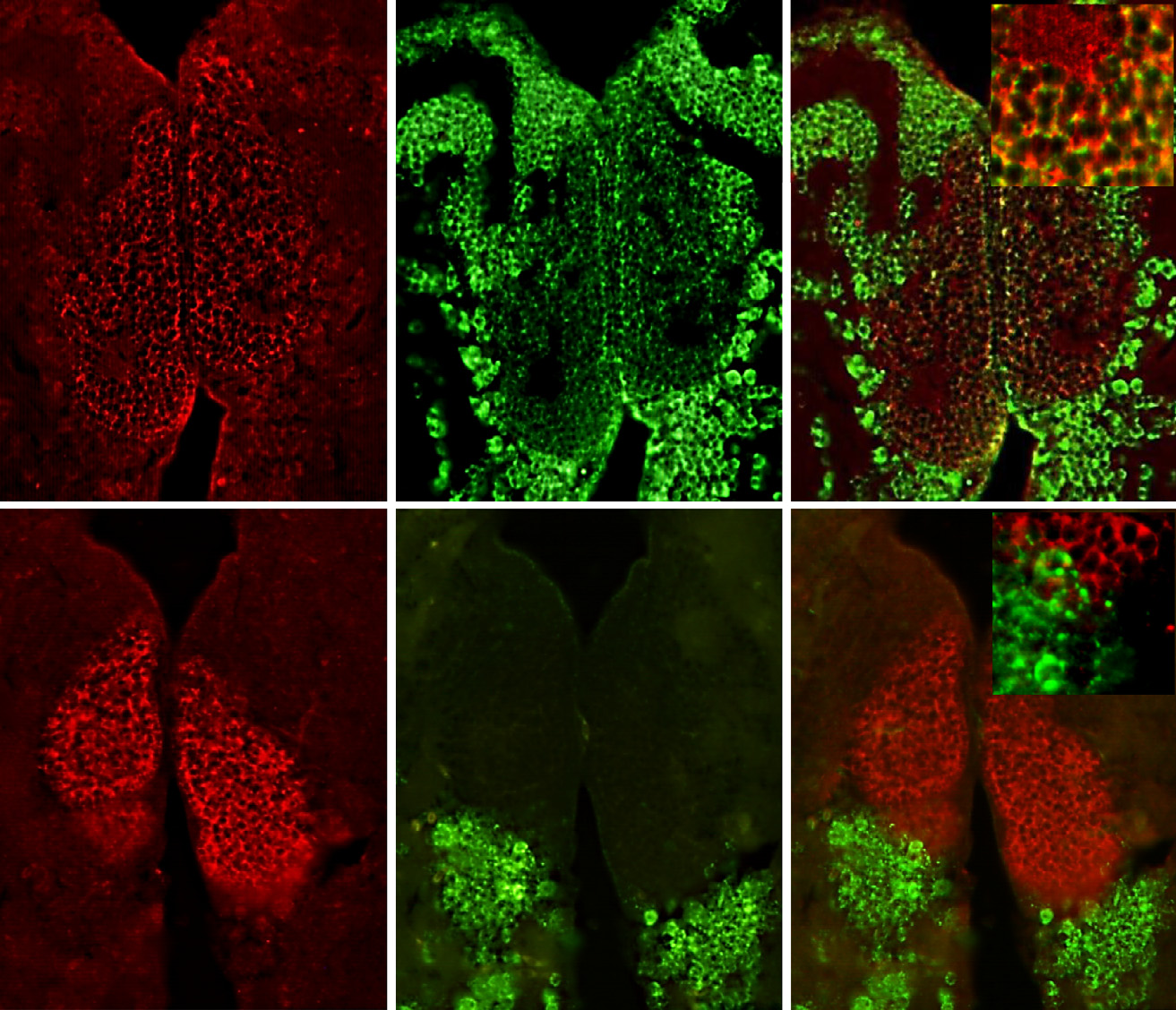
Dual-fluorescence labeling of kisspeptin 1 (Kiss1) and Kiss-R1 (red) in glutamatergic and GABAergic neurons (green) in the habenula. (a–c) Photomicrographs showing some populations of Kiss1-immunoreactive (-ir) cells in the vHb that co-localized with cells expressing slc17a6b mRNA. Some individual Kiss1-ir fibers originating from the vHb co-localized with slc17a6b mRNA (as seen in the magnified confocal inset representing the region enclosed by the orange-dotted box; 60× plus 1.5× optical zoom; N.A. = 1.4; z-step = 0.15 µm). No co-localization was observed between Kiss1-ir cells and gad2 mRNA-expressing cells. A: anterior thalamic nucleus. Scale bars, (a–f) 100 µm and inset c and f: 50 µm.