
|
Fig. 1
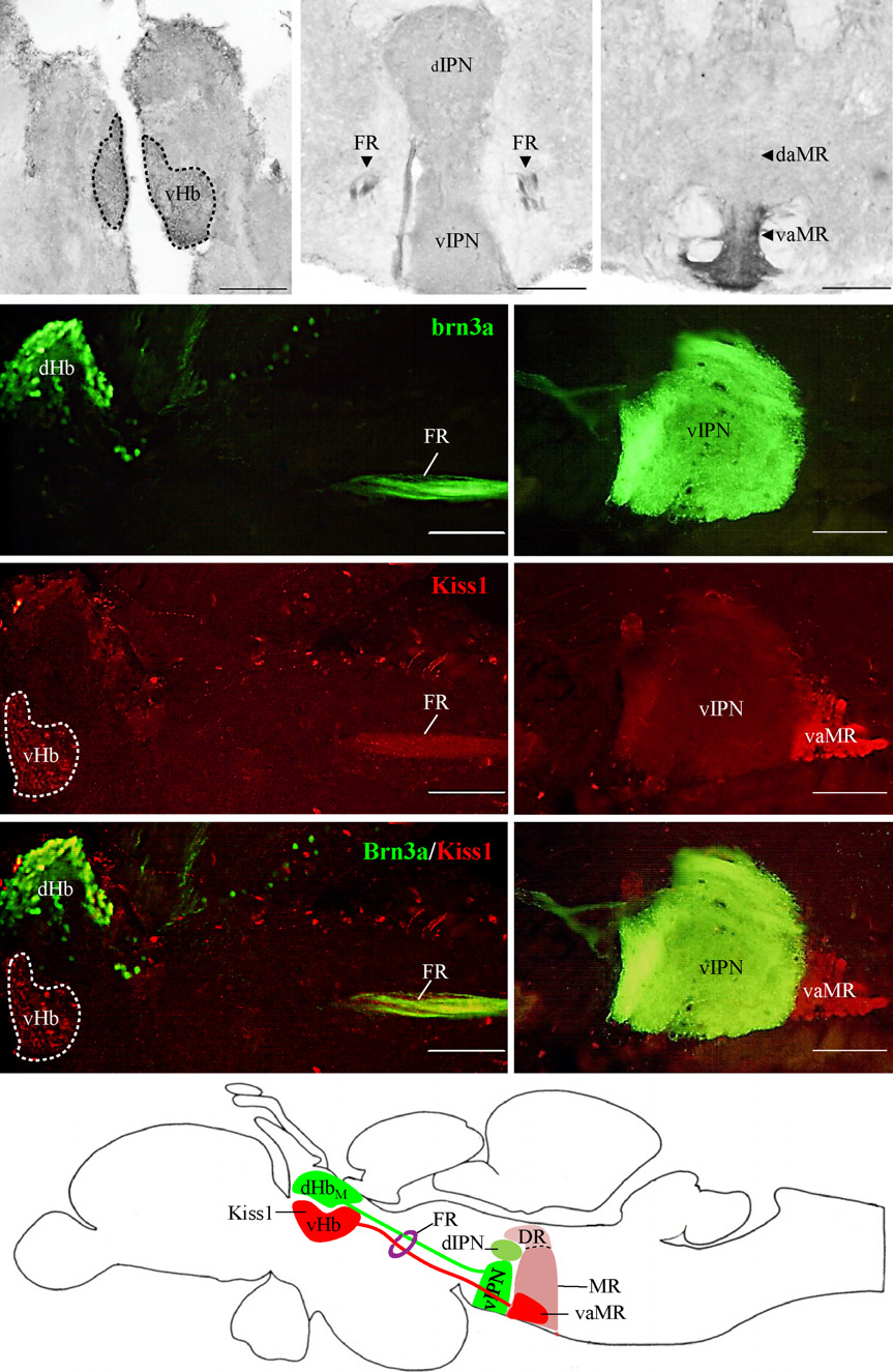
Kisspeptin 1 (Kiss1) projection in the zebrafish brain. (a–c) Kiss1-immunoreactive cells observed in the vHb (a) project through the fasciculus retroflexus (FR) (b) down to the vaMR (c). (d and e), Photomicrograph of sagittal section of Tg brn3a zebrafish expressing green fluorescent protein (GFP) in dHbM-vIPN pathway. (f–i) Kiss1-immunoreactive cells noted in vHb (red) and not in the dHb (green) with axonal projections coursing through the FR and terminating at the vaMR, a structure following the GFP-expressing vIPN. (j) Illustration depicts projections of dHbM and vHb through the FR to the vIPN and vaMR (a subregion of the MR) respectively as a representation of the sagittal images. Scale bars, 100 µm.