
Fig. 3
|
Fig. 3
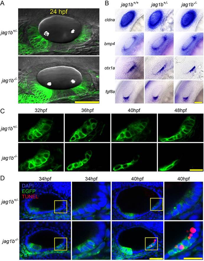
Jag1b is required for survival of the posterior prosensory cells. (A) Confocal images of the otic vesicle of embryos at 24hpf. Lateral views. Scale bar: 50µm. (B) Whole-mount in situ hybridization of otic marker genes at 24hpf. Scale bar: 20µm. (C) Time-lapse live imaging of the developing posterior prosensory domain. The posterior sensory cells are gradually lost in jag1b-/- embryos. Scale bar: 10µm. (D) TUNEL staining of embryos at 34hpf and 40hpf. At 40hpf, apoptosis signals are detected only in the posterior prosensory domain of jag1b-/- embryos. Dorsolateral views. Scale bars: 40µm (left) and 20µm (right). Anterior towards the left and dorsal upwards. Boxed regions are enlarged on the right.