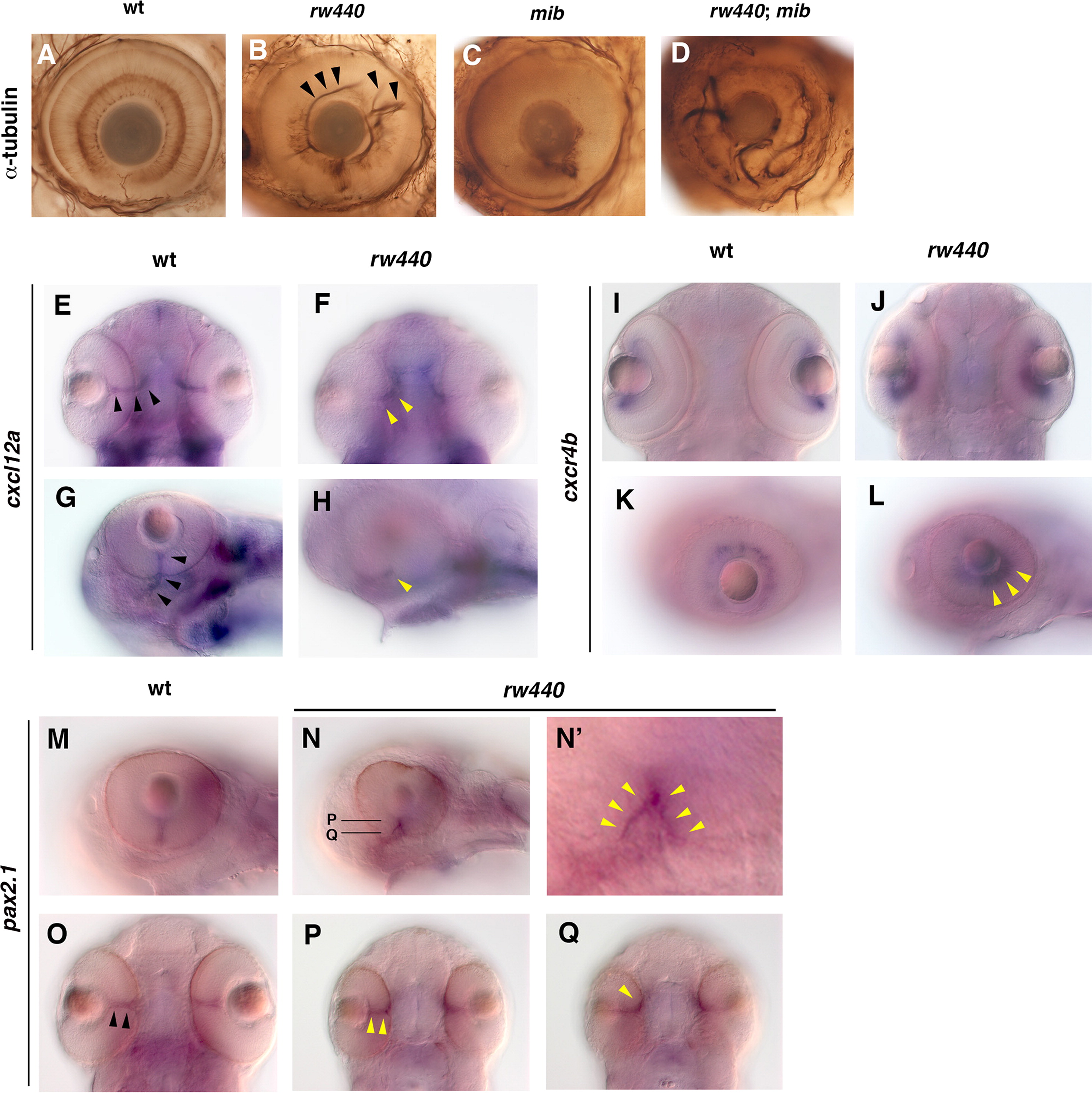
Fig. 8
The rw440 mutant shows defects in choroid fissure closure and spatial pattern of cxcl12a expression. (A–D) Anti-acetylated α-tubulin antibody labeling of wild-type (A), rw440 mutant (B), mib mutant (C), and rw440; mib double mutant (D) retinas. RGC axons fail to exit from the optic cup, but are misrouted within the optic cup in rw440 mutant retina (arrowheads, B). RGC axons are densely accumulated in the ventro-nasal retina but not misrouted in the mib mutant. RGC axons are more severely misrouted within the optic cup in the rw440; mib double mutant than in the rw440 mutant. (E–H) In situ hybridization of 60 hpf wild-type (E and G), and rw440 mutant (F and H) embryos with the cxcl12a mRNA probe. Ventral (EvF), and lateral (G and H) views are shown. In wild type, cxcl12a mRNA is expressed in the optic stalk, which is located inside the retina and the forebrain region (arrowheads, E and G). However, in the rw440 mutant, cxcl12a mRNA is only detected in the forebrain region (yellow arrowheads, F and H) but not inside the retina. (I–L) In situ hybridization of 60 hpf wild-type (I and K) and rw440 mutant (J and L) embryos with the cxcr4b mRNA probe. Ventral (I and J) and lateral (K and L) views are shown. In both wild type and rw440 mutant, cxcr4b mRNA is expressed in RGCs, although cxcr4b mRNA expression is stronger in the ventro-temporal region in the rw440 mutant (yellow arrowheads, L). (M–Q) In situ hybridization of 60 hpf wild-type (M and O), and rw440 mutant (N, P and Q) embryos with pax2.1 mRNA probe. (N′) High magnification of (N). In wild type, pax2.1 mRNA is expressed in the optic stalk (arrowheads, O). In the rw440 mutant, pax2.1 mRNA is expressed in a reverse V-shape along the ventral surface of the optic cup (yellow arrowheads, N′). (P and Q) Ventral view of deep (P) and superficial (Q) planes of the optic cup.
Reprinted from Developmental Biology, 394(1), Imai, F., Yoshizawa, A., Matsuzaki, A., Oguri, E., Araragi, M., Nishiwaki, Y., Masai, I., Stem-loop binding protein is required for retinal cell proliferation, neurogenesis, and intraretinal axon pathfinding in zebrafish, 94-109, Copyright (2014) with permission from Elsevier. Full text @ Dev. Biol.

