Fig. 3
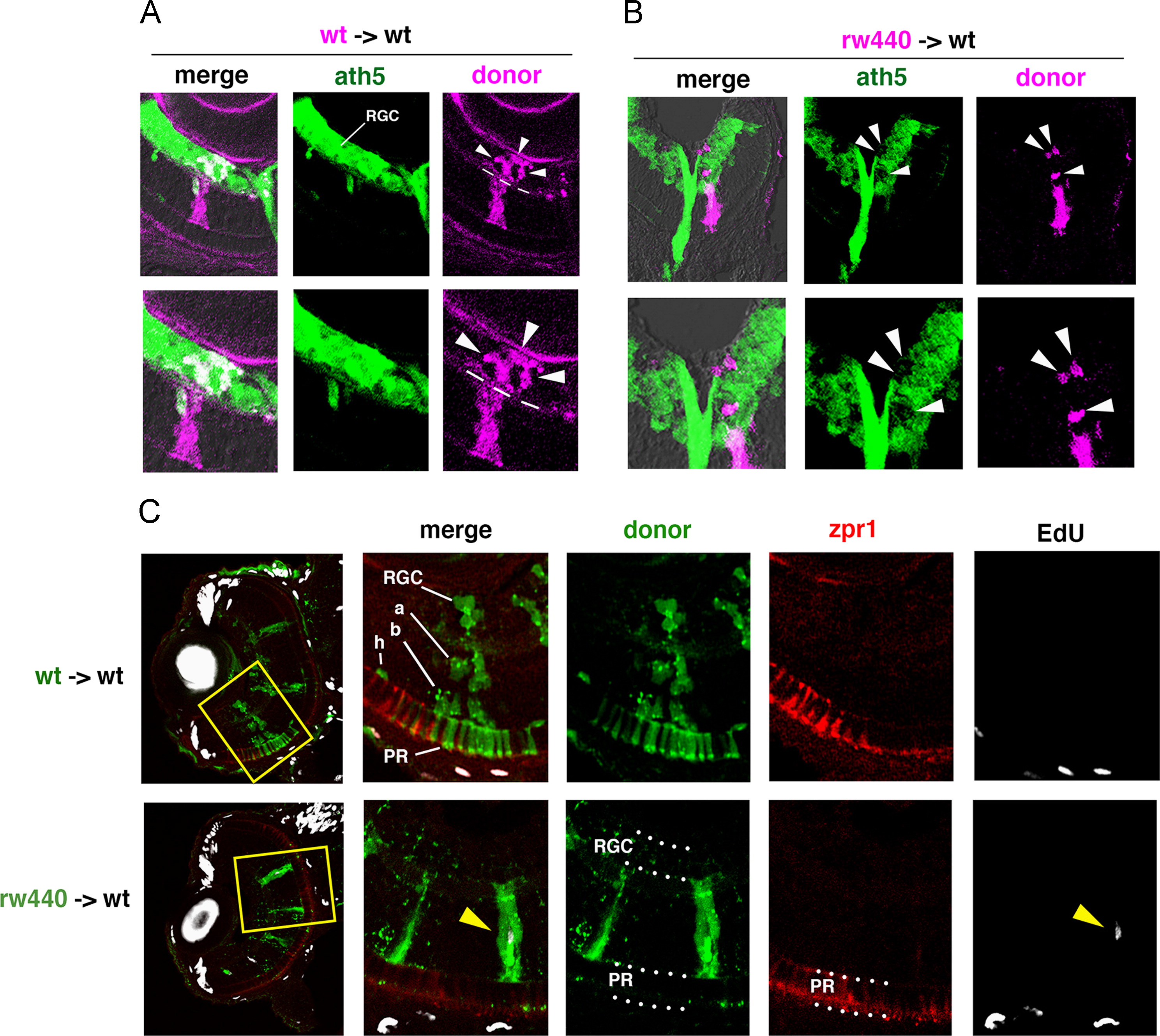
Cell transplantation experiments. (A) Transplantation of wild-type donor cells (magenta) into wild-type host retinas. Wild-type donor cells incorporated into RGL (arrowheads) express ath5:GFP (green) at 48 hpf. Bottom panels show high magnification. (B) Transplantation of the rw440 mutant donor cells (magenta) into wild-type host retinas. The rw440 mutant cells incorporated into the RGL (arrowheads) did not express ath5:GFP (green) at 48 hpf. Bottom panels show high magnification. (C) Seventy-two hpf wild-type host retinas into which wild-type donor cells (green, upper panels) or the rw440 mutant donor cells (green, lower panels) were incorporated. Retinal sections were labeled with EdU (white) and zpr1 antibody (red). Four sets of right panels are high magnification of the yellow squares shown in the left most panels. Wild-type donor cells into wild-type host retina differentiated into retinal ganglion cells (RGC), amacrine cells (a), bipolar cells (b), horizontal cells (h), and photoreceptors (PR) at 72 hpf. However, the rw440 mutant donor cells did not contribute to RGC and photoreceptors (PR) at 72 hpf, but displayed a progenitor-like, columnar morphology. Some of the rw440 donor mutant cells incorporated EdU (yellow arrowhead, lower right).
Reprinted from Developmental Biology, 394(1), Imai, F., Yoshizawa, A., Matsuzaki, A., Oguri, E., Araragi, M., Nishiwaki, Y., Masai, I., Stem-loop binding protein is required for retinal cell proliferation, neurogenesis, and intraretinal axon pathfinding in zebrafish, 94-109, Copyright (2014) with permission from Elsevier. Full text @ Dev. Biol.

