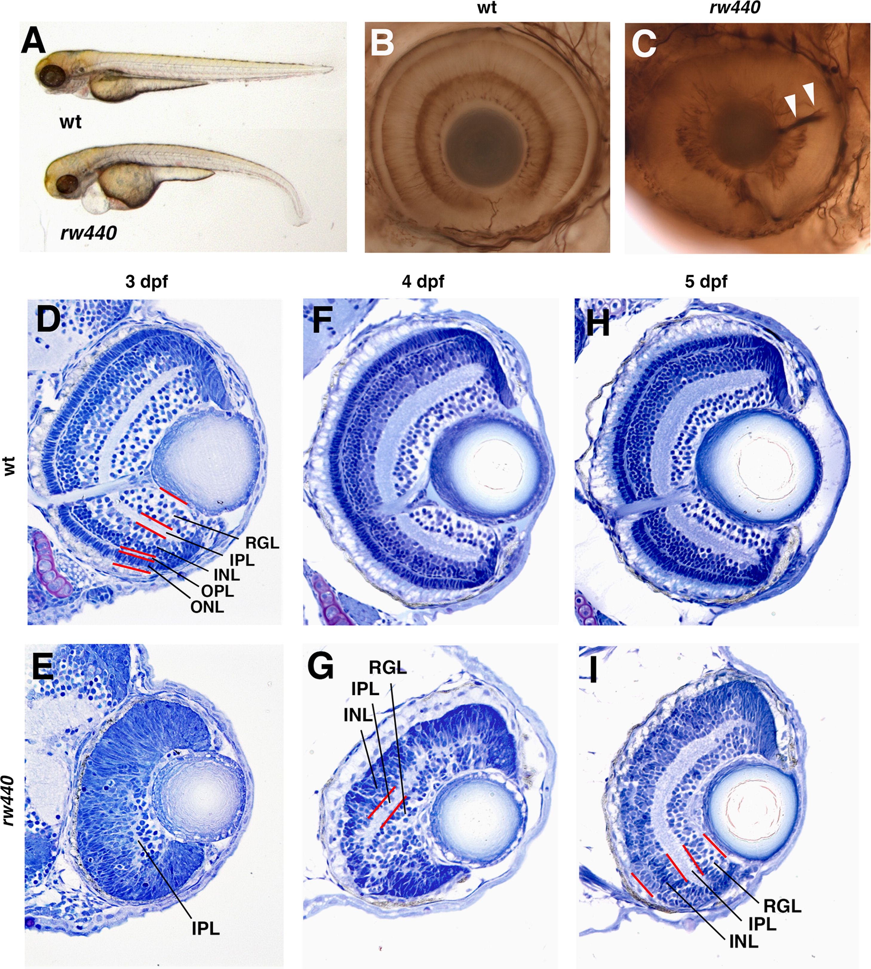
Fig. 1
Retinal lamination is delayed in the rw440 mutant. (A) Three dpf wild-type and rw440 mutant embryos. The rw440 mutant embryo shows a small eye, heart edema, and a curled-down body. (B and C) Labeling of 3 dpf wild-type (B) and rw440 mutant retinas (C) with anti-acetylated α-tubulin antibody. Retinal layers are visualized in the wild-type retina. However, retinal lamination is less developed, and retinal axons are misrouted inside the optic cup (arrowheads, C). (D and E) Sections of wild-type (D) and the rw440 mutant (E) retinas at 3 dpf. In wild-type retina, all layers are formed. However, in the rw440 mutant retina, retinal lamination is not observed, except a small IPL. (F and G) Sections of wild-type (F) and rw440 mutant (G) retinas at 4 dpf. In the rw440 mutant retina, the IPL becomes large, and the RGL and INL are distinct. (H and I) Sections of wild-type (H) and the rw440 mutant (I) retinas at 5 dpf. In the rw440 mutant retina, the RGL, IPL, and INL are more evident, but the ONL is absent. RGL, retinal ganglion cell layer; IPL, inner plexiform layer; INL, inner nuclear layer; OPL, outer plexiform layer; ONL, outer nuclear layer.
Reprinted from Developmental Biology, 394(1), Imai, F., Yoshizawa, A., Matsuzaki, A., Oguri, E., Araragi, M., Nishiwaki, Y., Masai, I., Stem-loop binding protein is required for retinal cell proliferation, neurogenesis, and intraretinal axon pathfinding in zebrafish, 94-109, Copyright (2014) with permission from Elsevier. Full text @ Dev. Biol.

