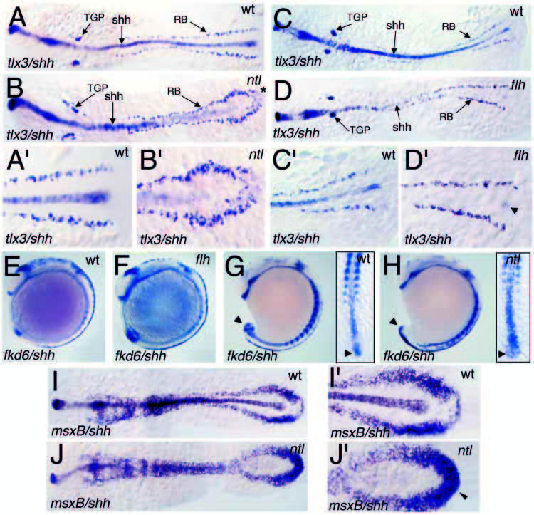
Fig. 5 Normal and expanded dorsal neural cell types in flh and ntl mutants, respectively. In 7-somite-stage embryos, rostral trunk regions of ntl mutants display normal numbers of tlx3- expressing RB precursors, while the caudalmost region, where shh expression is absent, exhibits an expansion in RB neurons (A,B, * marks the caudal region). (A′,B′) Enlargements of the posterior regions of A,B, showing the expanded RB cells in ntl mutants. In 10- somite-stage flh mutants, the RB cells and neural crest appear normal, even in regions where no shh-expressing midline cells are observed (C-F). (C′,D′) Enlargements of C,D showing weak shh expression in the tail bud of flh mutants. Caudal regions of ntl mutants display a mild expansion in the fkd6 neural crest domain at the 15-somitestage (G,H) and msxB expression in 7- somite-stage embryos (I,J). Arrowheads in G marks shh/fkd6 tail bud expression in wild type. Arrowheads in H indicate the expanded fkd6 expression domain in ntl mutants. (I′,J′) Enlargements of the posterior region of I,J, showing the loss of midline and tail bud shh expression in ntl mutants and expanded msxB expression. (A-J) Double in situ hybridizations including shh, except the insets in G and H where shh was omitted because tail bud shh expression obscures the fkd6 expression in flattened embryos.
Image
Figure Caption
Figure Data
Acknowledgments
This image is the copyrighted work of the attributed author or publisher, and
ZFIN has permission only to display this image to its users.
Additional permissions should be obtained from the applicable author or publisher of the image.
Full text @ Development

