Fig. 3
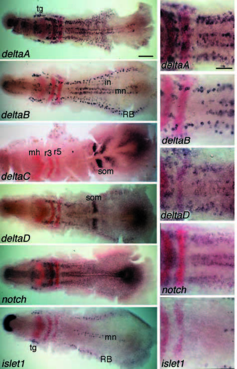
Fig. 3 Expression of deltaA, B, C and D, notch and islet1 at the 5- somite stage (11.7 hpf), shown by in situ hybridisation with a purple blue (NBT/BCIP) detection system; embryos have also been double labelled with probes against paxb and Krox20, using a Fast-Red detection system, to provide landmarks. On the right, enlargements of the posterior hindbrain and anterior spinal-cord region are shown. Embryos are flat-mounted; anterior is to the left. tg, trigeminal ganglion; mh, midbrain/hindbrain boundary; r3, r5, rhombomeres 3 and 5; mn, motor neurons; in, interneurons; RB, Rohon-Beard (sensory) neurons; som, prospective somite. Scale bars: 200 μm (whole embryos), 100 μm (details).

