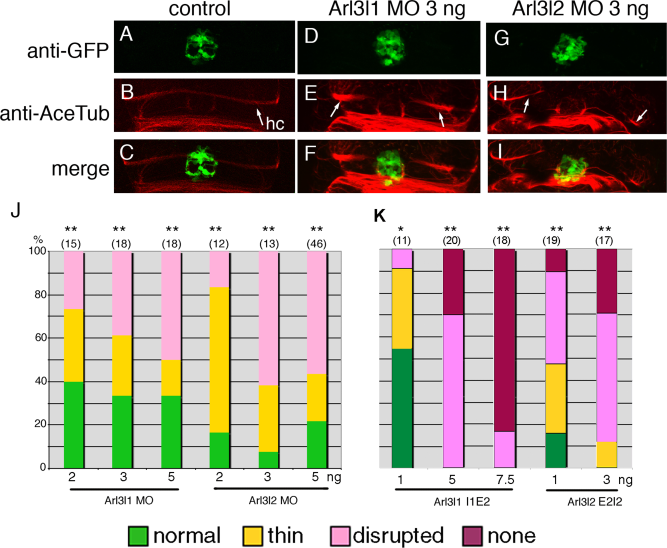
Fig. 2
Injection of arl3l1 and arl3l2 MO disrupts HC formation. Tg(AANAT2:eGFP) embryos were injected with control (A–C), arl3l1 (D–F, J), or arl3l2 (G–I, K) ATG MO, fixed at 54 hpf and stained with anti-GFP (green, pineal gland) (A, D, and G) and anti-acetylated tubulin (red, neurons) (B, E, and H). C, F, and I are merged images. Arrows indicate the HC (B, E, and H). Dose-dependence of the ATG MO effect (J). K: Splice MOs were injected into wild type embryos, which were stained for acetylated tubulin and classified as above. As the arl3l2 E2I2 MO was somewhat toxic, it was co-injected with 5 ng of p53 MO to suppress cell death (Robu et al., 2007). Experiments were compared to control HC, and statistical significance and number of embryos are indicated as specified in the legend to Figure 1.

