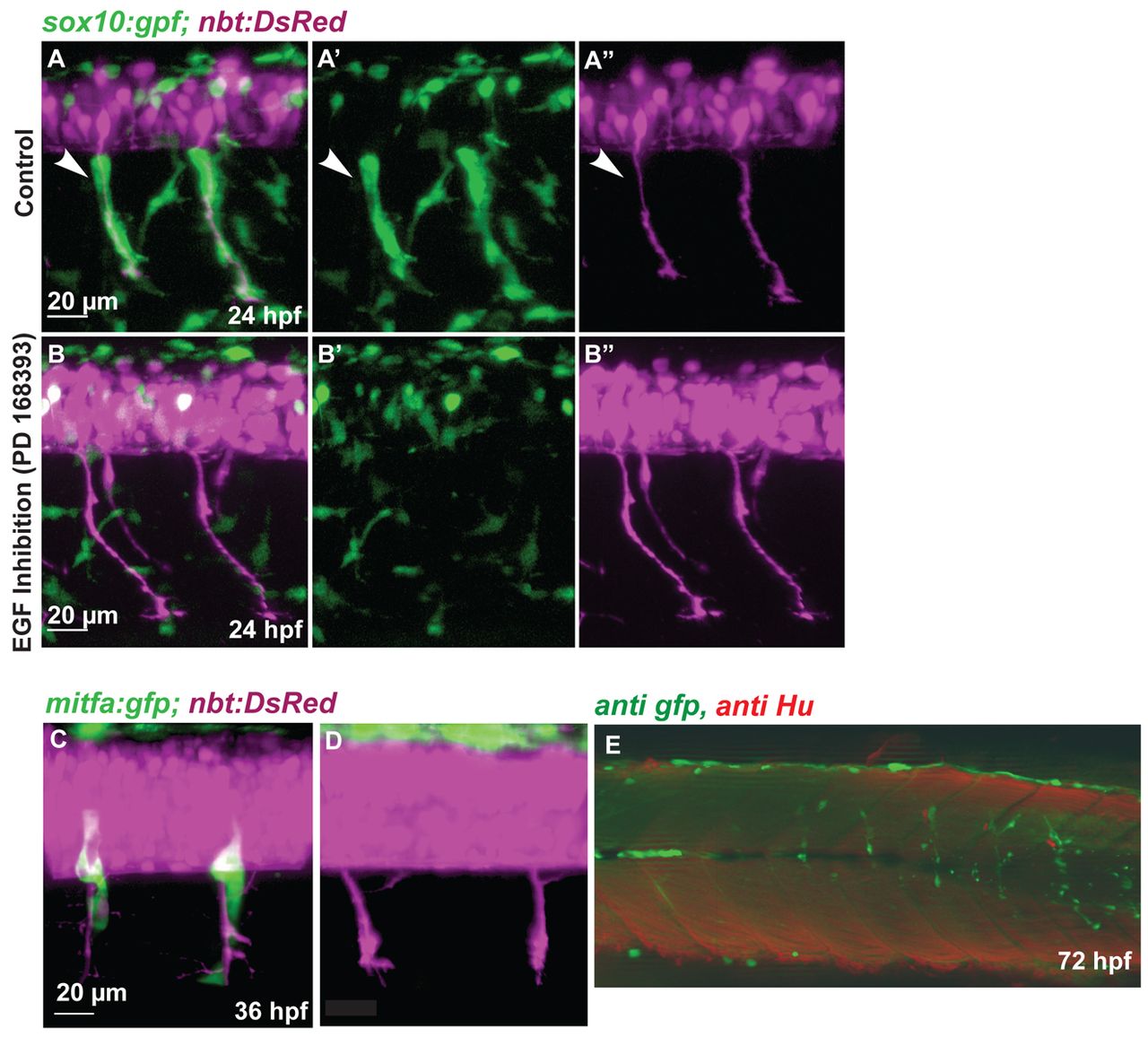
Fig. 6 NC migration along the ventromedial path is blocked by inhibition of ErbB receptors. (A-B′′) Tg(sox10:gfp; nbt:DsRed) zebrafish embryos at 24 hpf. (A,B) Red and green channel (merge); (A′,B′) green channel; (A′′,B′′) red channel. Medial NC cells (green) covering the primary motor axons (white arrowheads, A-A′′) are absent after treatment with the ErbB inhibitor PD168393 (B-B′′). (C,D) Confocal images of 36 hpf Tg(mitfa:gfp; nbt:DsRed) embryos. (C) Wild-type embryo. (D) Embryo treated with ErbB inhibitor at 16 hpf. (E) Tg(mitfa:gfp) embryos were injected with a double MO combination against mitfa and erbb3b. Larvae were stained at 8 dpf using anti-GFP (green) and anti-HU (red) antibody (white arrowheads). The association of DRGs (red) and GFP-positive cells was quantified (Table 1). In double mitfa and erbb3b knockdowns, of 82 metamers lacking HU positive cells only five (6%) develop a string of GFP-positive cells.
Image
Figure Caption
Figure Data
Acknowledgments
This image is the copyrighted work of the attributed author or publisher, and
ZFIN has permission only to display this image to its users.
Additional permissions should be obtained from the applicable author or publisher of the image.
Full text @ Development

