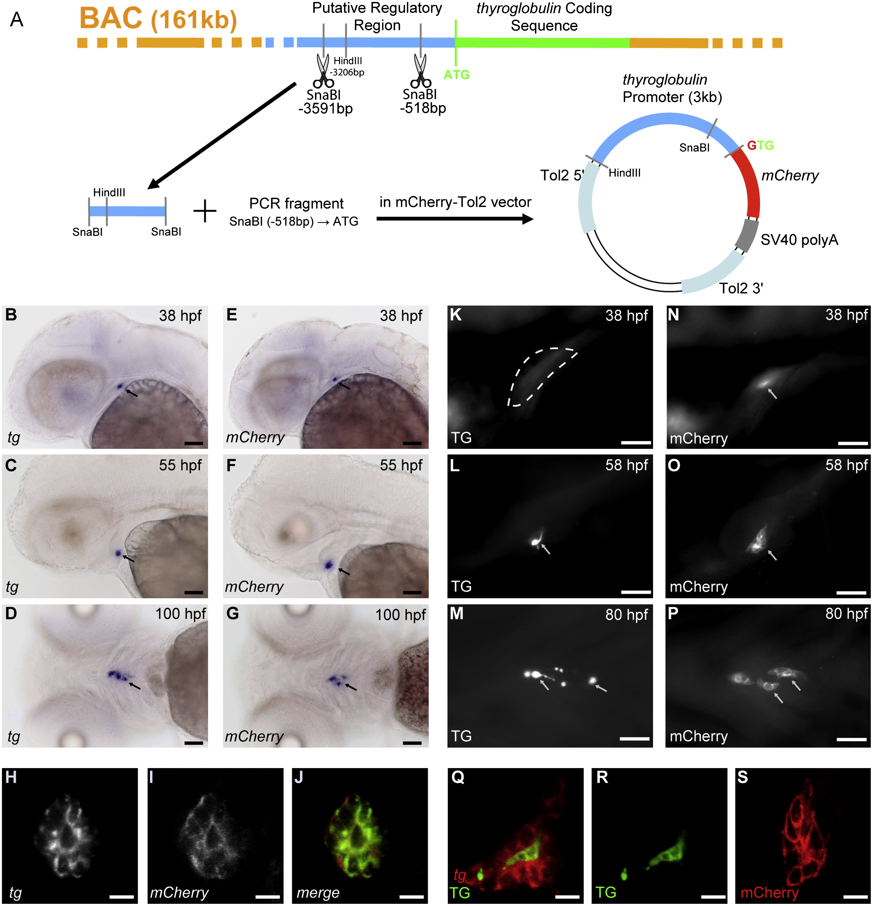
Fig. 1 Generation and characterization of tg(tg:mCherry) transgenic embryos. (A) Cloning strategy: A 3 kb region of the putative regulatory region of the zebrafish thyroglobulin coding sequence was isolated from a BAC clone by restriction with SnaBI. PCR was used to generate the remaining sequence upstream of the ATG, and a 3.2 kb DNA fragment was finally cloned upstream of the mCherry coding sequence. The final construct, flanked by Tol2 sites, was injected at the one-cell stage embryo. ((B)-(G)) mCherry mRNA expression in transgenic embryos recapitulates the thyroid-specific expression of endogenous tg mRNA as shown by whole-mount in situ hybridization. Arrows point to the thyroid. ((H)-(J)) Double fluorescent in situ hybridization (FISH) confirmed coexpression of mCherry and tg mRNA in all thyroid cells. Confocal images of a 55 hpf embryo are shown. ((K)-(P)) Whole-mount immunofluorescence (IF) of transgenic embryos revealed mCherry protein expression ((N)-(P)) as early as 36/37 hpf, well before thyroglobulin (TG) protein ((K)–(M)) becomes detectable around 55 hpf. The encircled area in panel (K) marks the thyroid region which lacks staining for TG protein at 38 hpf. Arrows point to the thyroid. ((Q)-(S)) TG staining is limited to the colloid (see (Q) and (R)) whereas mCherry IF staining labels all thyroid cells (see (S)). Panels (Q) and (R) show a confocal section of the thyroid of a 60 hpf embryo after combined staining for tg mRNA (FISH) and TG protein (IF). Panel (S) shows a confocal section of the thyroid of a 55 hpf embryo after IF staining for mCherry. Ventral views are shown in (D), (G), (M) and (P), all other images show lateral views. All embryos shown are oriented with anterior to the left. Scale bar: 50 μM in (B)-(G), (K)-(P) and 10 μM in (H)-(I), (Q)-(S).
Reprinted from Developmental Biology, 372(2), Opitz, R., Maquet, E., Huisken, J., Antonica, F., Trubiroha, A., Pottier, G., Janssens, V., and Costagliola, S., Transgenic zebrafish illuminate the dynamics of thyroid morphogenesis and its relationship to cardiovascular development, 203-216, Copyright (2012) with permission from Elsevier. Full text @ Dev. Biol.

