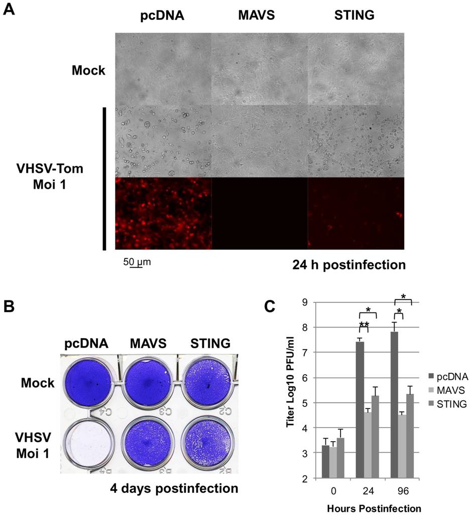
Fig. 2 Zebrafish STING is a strong antiviral protein.
EPC cells were transfected with 2 μg of pcDNA-STING, and pcDNA-MAVS or an empty vector (pcDNA) as positive and negative controls, respectively. At 48 h posttransfection, EPC cells were infected with a recombinant rVHSV-Tom expressing the tdTomato fluorescent protein at an MOI of 1 and incubated at 15°C. Cell monolayers were visualized under a UV-visible light microscope at 24 h postinfection (A) and then stained with crystal violet 4 days postinfection (B). The culture supernatants from cells infected with rVHSV-Tom were collected at 0, 24 and 96 h postinfection and the viral titer was determined by plaque assay on EPC cells (C). Each time point was represented by three independent experiments, and each virus titration was done in duplicate. Means are shown. The standard errors were calculated and the error bars are shown. Asterisks indicate significant difference (*p<0.01 and **p<0.001) as determined by Student′s t test.

