Image
Figure Caption
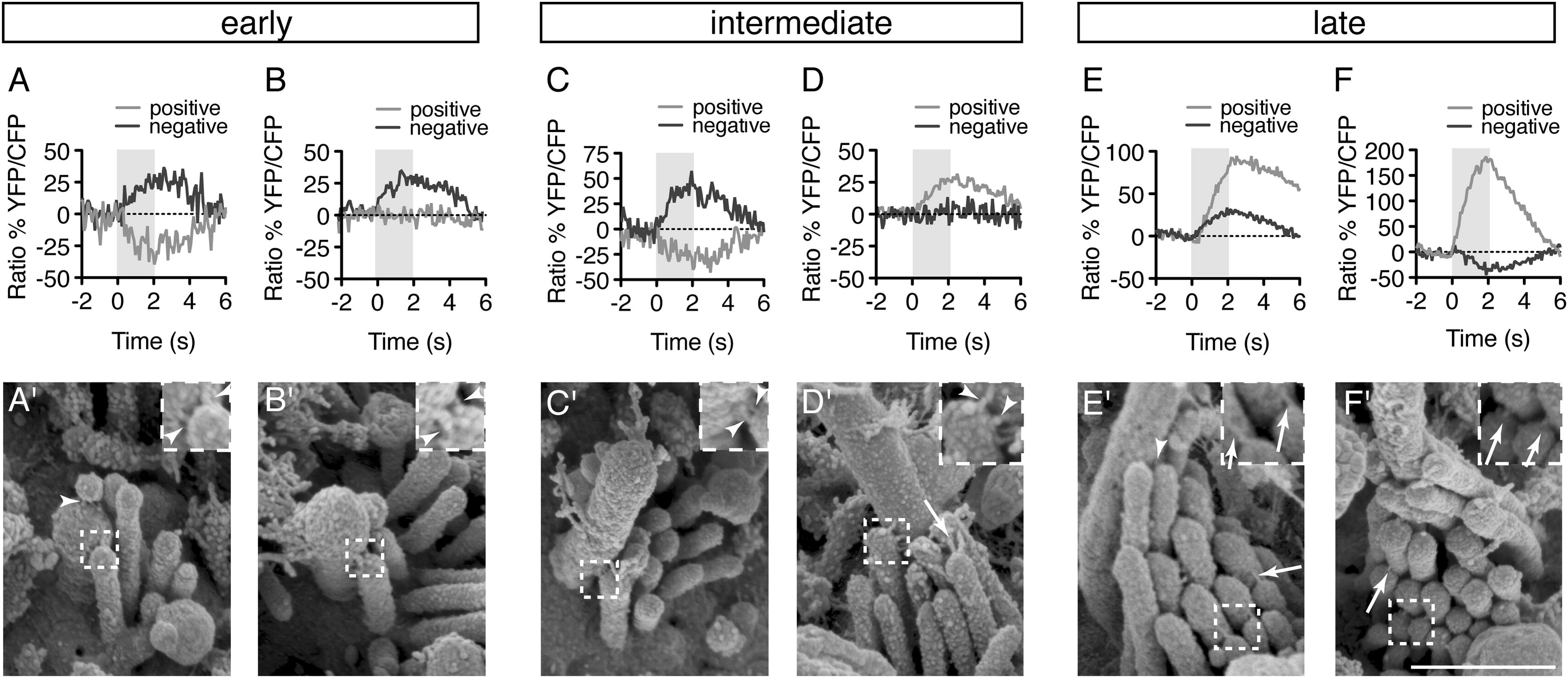
Fig. 5 Correlative Calcium Imaging and SEM Reveal a Change in Link Architecture during Development(A–F) Calcium responses were measured in response to positive and negative hair-bundle deflections from cells at each stage of bundle development.(A′–F′) SEM images of the hair bundles from hair cells represented in (A)–(F).All images were taken at 3 dpf. Arrowheads indicate kinocilial links; arrows indicate tip links. Scale bar, 500 nm. Inset is 2× magnification. For additional link examples see also Figure S3.
Acknowledgments
This image is the copyrighted work of the attributed author or publisher, and
ZFIN has permission only to display this image to its users.
Additional permissions should be obtained from the applicable author or publisher of the image.
Reprinted from Developmental Cell, 23(2), Kindt, K.S., Finch, G., and Nicolson, T., Kinocilia mediate mechanosensitivity in developing zebrafish hair cells, 329-341, Copyright (2012) with permission from Elsevier. Full text @ Dev. Cell

