Fig. 6
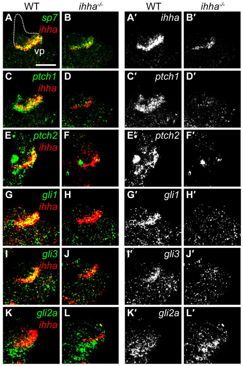
Fig. 6 Hh signaling is regionally localized. Confocal sections showing Hh pathway gene expression at 5 dpf by whole-mount in situ hybridization. Images show expression of genes relative to ihha (A-L) and a separated channel with individual gene expression (A′-L′) from respective images. Images are lateral views with dorsal upwards and anterior towards the left. (A,A′) ihha is co-expressed with sp7 and specifically demarcates the vp edge. Other Op edges deduced by DIC are shown with a broken line. (B,B′) sp7 expression appears similar to wild type in ihha mutants, although the domain may be slightly reduced, reflecting the diminished vp edge length. ihha expression is reduced in ihha mutants. (C,C′) ptch1 is expressed ventral to vp edge osteoblasts and is also co-expressed with ihha along the vp edge. (D,D′) Expression of ptch1 is lost in ihha mutants. (E-F2) ptch2 expression resembles that of ptch1. (G-J′) gli1 and gli3 are co-expressed with ihha and their expression is decreased in ihha mutants. (K-L′) gli2a is expressed just ventral to the vp edge and its expression is unaltered in ihha mutants. Scale bar: 50 μm.

