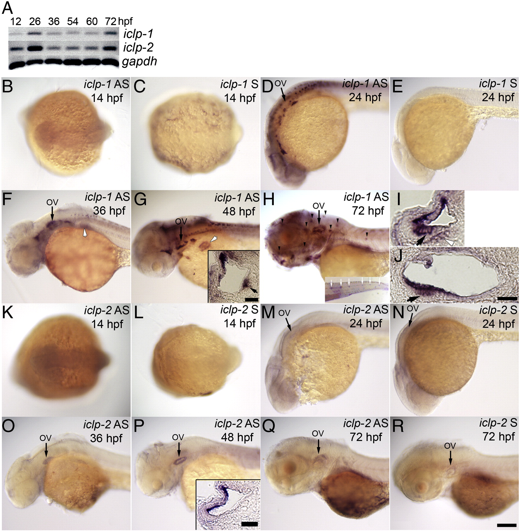
Fig. 3
Expression patterns of embryonic iclp1 and iclp2. (A) RT-PCR results of iclp1 and iclp2 transcripts from whole embryos of 12, 26, 36, 54, 60, and 72 hpf. (B–H, K–R) Whole mount ISH with iclp1 (B–H) or iclp2 (K–R) probes. (B, C, K, L) Dorsal view of 14-hpf embryos; (D–H, M–R) Lateral view of (D, E, M, N) 24-hpf embryos; (F, O) 36-hpf embryos; (G, P) 48-hpf embryos; (H, Q, R) 72-hpf larvae. (B, D, F, G, H) with iclp1 antisense probe; (C, E) with iclp1 sense probe; (K, M, O, P, Q) with iclp2 antisense probe; (L, N, R) with iclp2 sense probe. (I, J) iclp1 labeling of 48 hpf embryo with transverse (I) and sagittal (J) sections. Arrows in (I) and (J) indicate hair cells in the anterior macula. Insets in (G) and (P) show transverse sections of the ear; Inset (G) inset shows a section through the posterior macula; small arrow in inset (G) indicates hair cells in the sensory patches. Inset (H) shows a tail with posterior LL neuromast staining. OV: otic vesicle. Arrowheads: anterior LL neuromasts; white arrows: posterior LL neuromasts; white arrows with black outlines: pectoral fin bud; white arrowhead with black outline: statoacoustic ganglion (SAG). Scale bar: 100 μm for (B–H) and (K–R); 20 μm for insets, (I) and (J).
Reprinted from Developmental Biology, 363(1), Shen, Y.C., Thompson, D.L., Kuah, M.K., Wong, K.L., Wu, K.L., Linn, S.A., Jewett, E.M., Shu-Chien, A.C., and Barald, K.F., The cytokine macrophage migration inhibitory factor (MIF) acts as a neurotrophin in the developing inner ear of the zebrafish, Danio rerio, 84-94, Copyright (2012) with permission from Elsevier. Full text @ Dev. Biol.

