
|
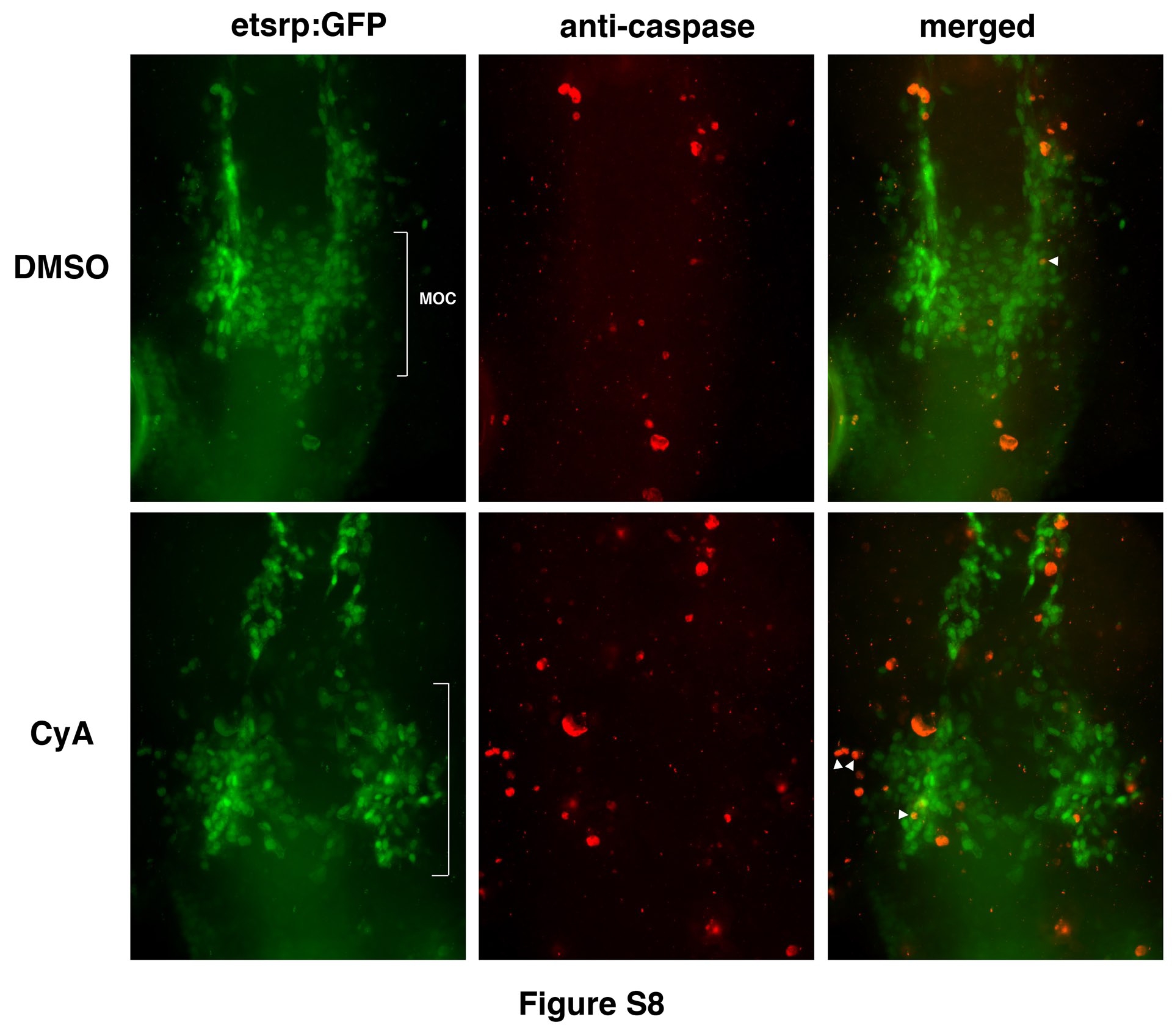
Fig. S8
CyA treatment does not result in increased apoptosis among endocardial progenitors.
Overlay (right panels) of etsrp:GFP (left) and anti-caspase staining (middle) at the 14-15-somite stages. Maximal projection images are shown. Note that multiple apoptotic and etsrp:GFP cells are in different Z-planes from cells although they may appear to overlap in the projection image. Overlapping staining is shown in white arrowheads. Anti-caspase stained cells were quantified in the MOC region (while brackets). As analyzed by Student’s t-Test, the average numbers and standard deviation of anti-caspase stained etsrp:GFP cells per embryo were 4.1±3.2 (n=11) and 6.0±3.9 (n=8) in DMSO control and CyA-treated samples, respectively, which is not statistically different (p=0.25).
Reprinted from Developmental Biology, 361(2), Wong, K.S., Rehn, K., Palencia-Desai, S., Kohli, V., Hunter, W., Uhl, J.D., Rost, M.S., and Sumanas, S., Hedgehog signaling is required for differentiation of endocardial progenitors in zebrafish, 377-91, Copyright (2012) with permission from Elsevier. Full text @ Dev. Biol.