Fig. S3
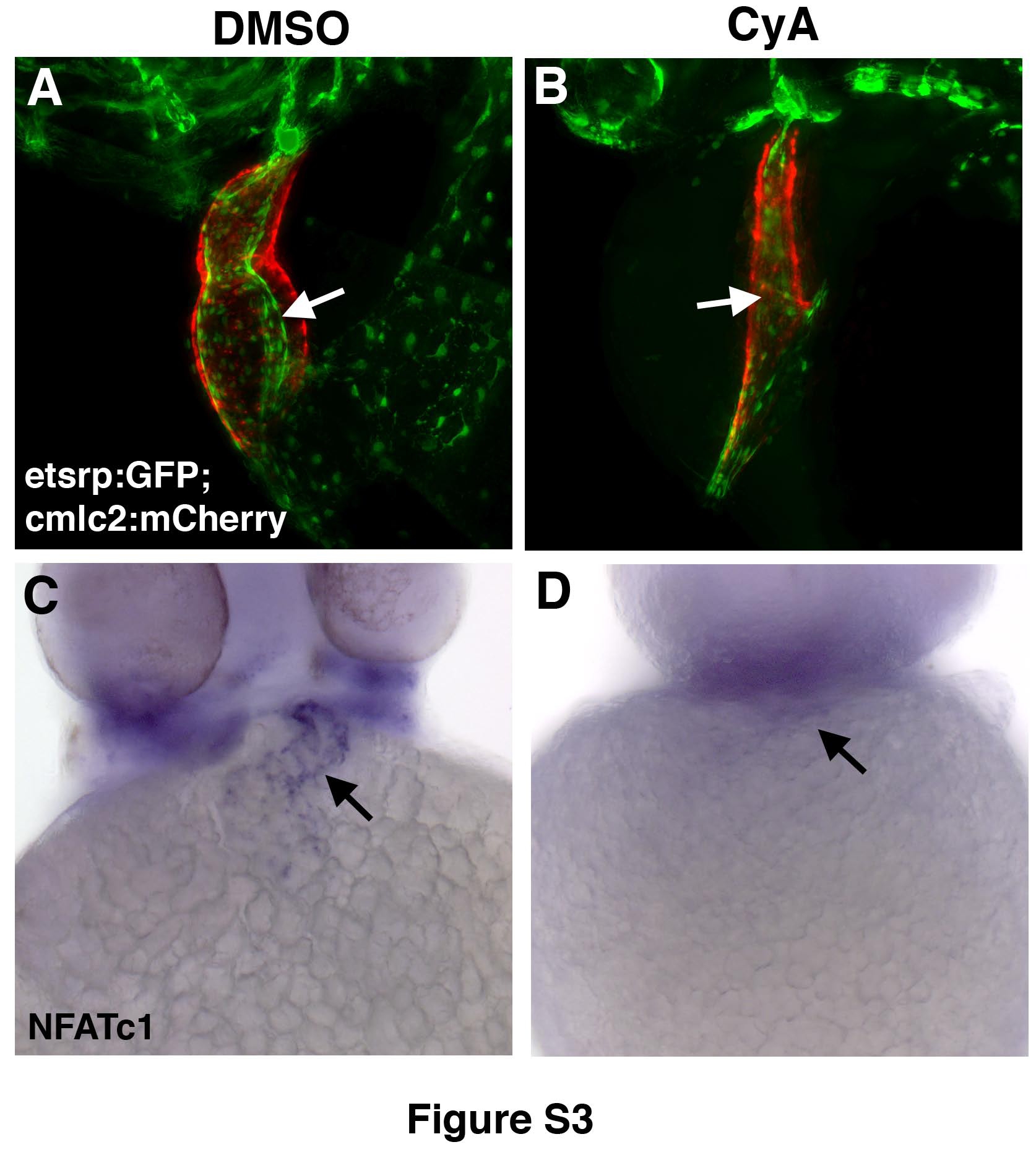
Reduced endocardium is present in CyA-treated embryos at 48hpf while NFATc1 expression is absent.
(A,B) Endocardium and myocardium visualized by etsrp:GFP (green) and cmlc2:mCherry (red) expression in live DMSO control (A) and CyA-treated embryos (B). Note the presence of reduced myocardium and endocardium and the absence of heart looping in CyA-treated embryos. (C,D) Endocardial NFATc1 expression is absent in CyAtreated embryos (D) as compared to DMSO-treated controls (C). All panels, ventral view of whole-mounted embryos, anterior is to the top. (A,B) Maximal projection images were obtained using compound fluorescence microscopy (Zeiss, AxioImager) and subjected to 3Ddeconvolution (AutoQuant software package).
Reprinted from Developmental Biology, 361(2), Wong, K.S., Rehn, K., Palencia-Desai, S., Kohli, V., Hunter, W., Uhl, J.D., Rost, M.S., and Sumanas, S., Hedgehog signaling is required for differentiation of endocardial progenitors in zebrafish, 377-91, Copyright (2012) with permission from Elsevier. Full text @ Dev. Biol.

