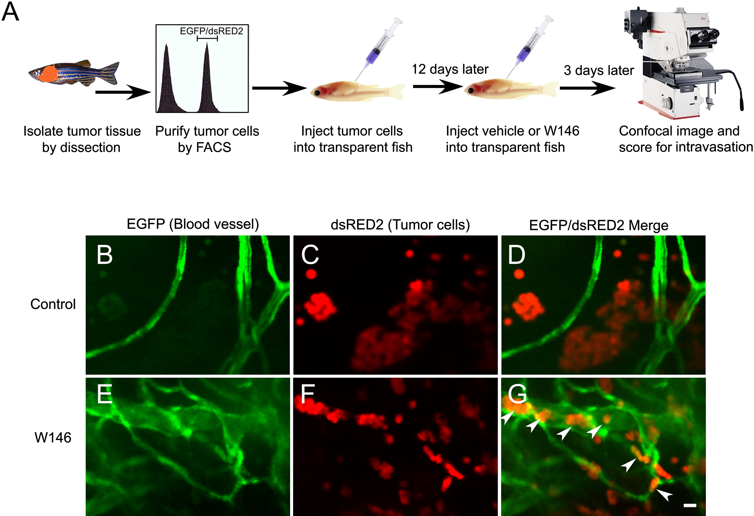
Fig. 8 The Selective S1P1 Antagonist W146 Promotes Intravasation of Bcl-2-Overexpressing T-LBL Cells In Vivo
(A) Schematic drawing of the experimental strategy.
(B–G) Confocal images of EGFP-labeled blood vessels (B and E), dsRED2-labeled lymphoma cells (C and F), and the merged images of a vehicle-treated (D; n = 29) and a W146-treated transplanted animal (G; n = 18) demonstrate that W146 treatment promotes intravasation of bcl-2-overexpressing lymphoma cells (arrowheads) in vivo (cf. G to D). Note that W146 treatment also inhibited the in vivo formation of lymphoma cell aggregates (cf. F to C).
Scale bar for (B)–(G), 10 μM.
Reprinted from Cancer Cell, 18(4), Feng, H., Stachura, D.L., White, R.M., Gutierrez, A., Zhang, L., Sanda, T., Jette, C.A., Testa, J.R., Neuberg, D.S., Langenau, D.M., Kutok, J.L., Zon, L.I., Traver, D., Fleming, M.D., Kanki, J.P., and Look, A.T., T-lymphoblastic lymphoma cells express high levels of BCL2, S1P1, and ICAM1, leading to a blockade of tumor cell intravasation, 353-366, Copyright (2010) with permission from Elsevier. Full text @ Cancer Cell

