
|
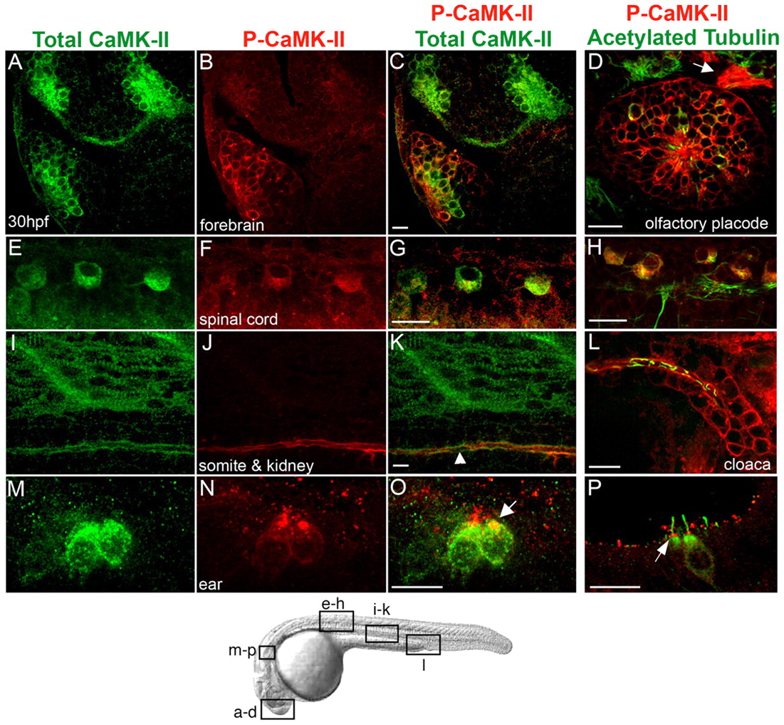
Fig. 1
Localization of activated and total CaMK-II in 30 hpf zebrafish embryos. (A-P) Embryos were immunolabeled for total CaMK-II (A,E,I,M) and P-CaMK-II (B,F,J,N) and imaged laterally in the forebrain (A-C), spinal cord cell bodies (E-G), muscle sarcomeres and the pronephric kidney (I-K), and embryonic ear (M-O) were examined (see diagram below). P-CaMK-II colocalizes with acetylated tubulin in some cells and axons in a frontal view of the olfactory placode (D, arrow) and in spinal cord cell bodies (H), but is undetectable in spinal cord axons. P-CaMK-II localizes at the apical surface of pronephric ductal cells (K, arrowhead), at all surfaces of cloacal cells (L) and at the base of the hair cell kinocilium (O, P, arrows). Scale bars: 10 μm