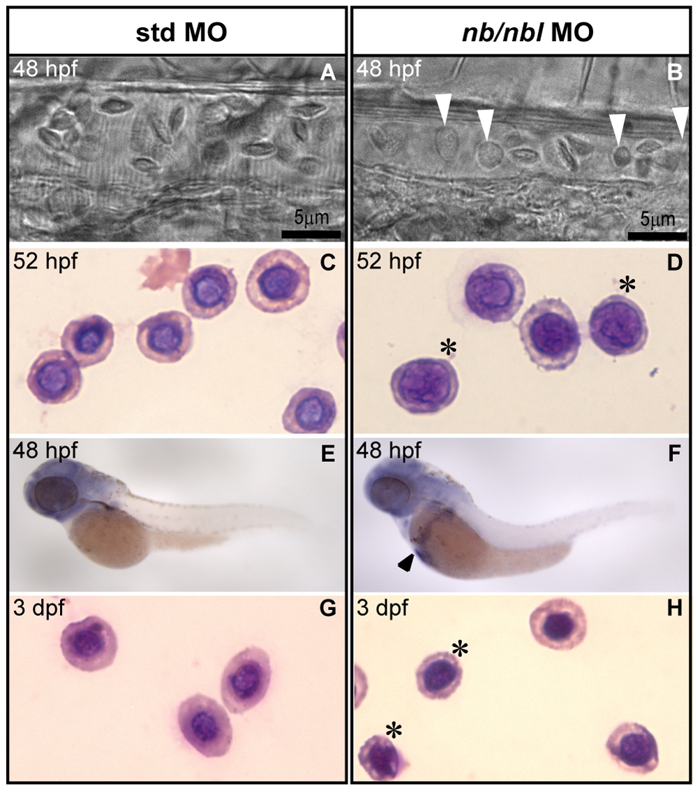
Fig. 5 Erythroblasts fail to differentiate into erythrocytes in apparently unaffected nb/nbl morphants.
A–B. Bright-field microscopy of blood cells in the caudal arteries of living 48 hpf std MO embryos (A) and nb/nbl morphants (B). nb/nbl MO-injected embryos with no apparent hematopoietic defects show the presence of abnormal-shaped blood cells (white arrowheads) in the blood flow (B). C–D, G–H. Wright-Giemsa staining of circulating embryonic red blood cells from controls and nb/nbl morphants at 52 hpf (C, D) and 3 dpf (G, H). Erythroid cells of apparently unaffected nb/nbl MO injected embryos were larger, showed a large nucleus and had more basophilic cytoplasm indicating the presence of maturation defects (D, H). Asterisks indicate representative erythroid cells with maturation defects. E–F. WISH using gata1 on 48 hpf std MO (E) and nb/nbl MO-injected embryos (F). gata1 expression persists in red blood cells of nb/nbl morphants revealing the presence of immature red cells (black arrowhead).

