Fig. 1
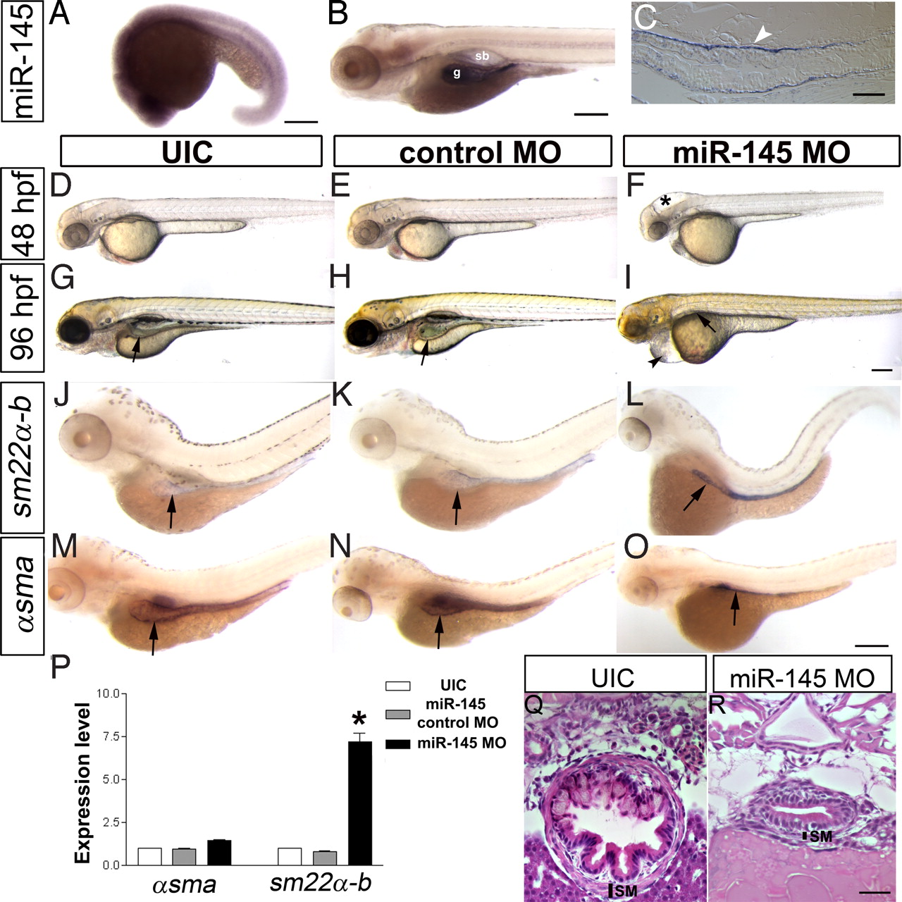
miR-145 expression pattern and loss of function phenotype. (A) Lateral view of whole-mount in situ expression of miR-145 at 16–19S shows ubiquitous expression. (B) Later at 96 hpf, miR-145 is expressed in the gut (g) and swimbladder (sb) in wild-type embryos. (C) Longitudinal section of 96 hpf embryo shows miR-145 expressed strongly in the gut smooth muscle cells (white arrowhead) and weakly in gut epithelium. (D–F) Animals injected with miR-145 MO have hydrocephalus in the hind brain ventricle (*) at 48 hpf. (G–I) At 96 hpf, miR-145 morphants have severe pericardial edema (black arrowhead), and an underdeveloped gut and swimbladder (black arrow). In situ hybridization staining of αsma (J–L) and sm22α-b (M–O) in 96 hpf embryos shows an increase of sm22α-b expression but no change of αsma in the miR-145 morphant gut. Gut morphology in the miR-145 morphant is unlooped as compared to the broader tube-like morphology found in UIC and miR-145 control MO embryos. Arrows mark the gut. (P) qPCR of αsma and sm22α-b in miR-145 morphants at 48 hpf. (Q and R) Histology of 96 hpf zebrafish gut. miR-145 morphant shows a thinner layer of SMCs (SM) and lack of villi in the epithelial layer as compared to UIC embryos. (Scale bars, 200 μm except for C, Q, and R, which are 50 μm.)

